What to Expect During a Full Body MRI: Prep & Process
You book the scan because you want clarity. Not because you feel sick, but because you'd rather understand your health before symptoms force the conversation. Then a second thought arrives just as quickly. What happens during a full body MRI, and what will it feel like when the day comes?
Individuals who schedule this kind of imaging are thoughtful, health-conscious, and a little uncertain at the same time. They're comfortable investing in prevention, yet they still wonder about the machine, the noise, the preparation, and the possibility of hearing about something unexpected. Those questions are reasonable.
A full body MRI can be an advanced, informative tool when it's used with clinical judgment. It's also a procedure that's often misunderstood. The imaging itself is straightforward, but the meaning of what it finds can be more nuanced than many patient-facing guides suggest. If you're considering this step, it helps to know the experience from beginning to end, and to understand why imaging quality and interpretation matter as much as the scan itself. For a broader look at how this fits into preventive care, see whole-body MRI for longevity and integrated personalized medical care.
Embarking on Your Proactive Health Journey
A common scenario looks like this. You've had your routine labs. You try to exercise consistently. You pay attention to sleep, recovery, and family history. Yet there's still a quiet sense that standard screening only shows part of the picture. A full body MRI feels like the next logical step, but also like stepping into unfamiliar territory.
That mix of curiosity and caution is healthy. MRI technology can produce remarkably detailed images of the body without ionizing radiation. It's non-invasive, and for screening purposes it often doesn't require contrast. Still, the experience is different from a routine office visit. You'll spend time in a highly technical environment, and you may hear terms that aren't part of everyday conversation.
Why many patients choose this scan
Some people want a baseline. Others want to look more closely because of family history, prior health concerns, or a general commitment to prevention. The strongest reason to pursue imaging is not fear. It's informed ownership.
A helpful analogy is this. Blood work is like reading a dashboard. A full body MRI is more like walking through the engine room with a flashlight. You may not need it at every stage of life, and it doesn't replace a skilled physician's judgment, but it can reveal structural details that routine screening won't show.
A good MRI experience doesn't begin when you enter the scanner. It begins when you understand what the scan can show, what it can't, and how the results will be interpreted.
Not every scan creates the same kind of clarity
Understandably, many readers get confused. The phrase “full body MRI” sounds uniform, as though every facility performs the same study in the same way and interprets it through the same lens. That isn't how advanced imaging works.
Protocol design matters. Radiology review matters. Clinical context matters. When those pieces are disconnected, patients may leave with more ambiguity than understanding. When they're integrated, the same scan can feel far more useful and far less emotionally disruptive.
If you're wondering what to expect during a full body MRI, the practical answer is simple. You'll prepare carefully, lie still for a defined period, hear a series of loud rhythmic sounds, and then wait for a detailed interpretation. The more important answer is that the quality of your experience depends on how thoughtfully the process is managed from the start.
How to Prepare for Your Full Body MRI Scan
Preparation is usually simple, but it's important. Small details can affect both safety and image quality. A little planning helps the appointment feel calm instead of rushed.

What to do before you arrive
Use this short checklist in the day or two before your appointment.
Choose metal-free clothing. Soft, comfortable garments are easiest. Avoid zippers, snaps, metallic threads, underwire, and decorative hardware.
Remove jewelry and wearable devices. Rings, watches, earrings, hearing devices, and fitness trackers all need attention before scanning.
Follow any food instructions exactly. Some centers ask you to fast for a period before the scan. If your imaging team gives you instructions, follow those rather than guessing.
Review your medications in advance. If you take prescription medications, ask whether you should continue them as usual.
Complete the screening questionnaire carefully. This is not routine paperwork. It's a safety document.
For a closer look at the technology side, how an AI full body MRI works explains why preparation and protocol design matter so much.
Why metal matters so much
MRI uses powerful magnetic fields and radio waves to generate images. It does not use ionizing radiation, which is one reason many patients find it appealing compared with X-ray based imaging. But that same magnetic environment is why metal has to be taken seriously.
External metal can interfere with image quality. Internal metal can raise more complex questions depending on the type of implant or device. Pacemakers, clips, stimulators, joint hardware, ports, or prior surgical materials all need to be disclosed before the appointment. The safest approach is complete transparency, even if you're unsure whether something is relevant.
Practical rule: if it's implanted, worn, clipped, pierced, magnetic, or metallic, mention it.
A few practical expectations
A full-body MRI scan typically lasts between 60 to 90 minutes, insurance rarely covers it for asymptomatic individuals, and the American Cancer Society cautions that a negative scan may create a false sense of security, as described in Sermo's review of full-body MRI screening. That means preparation isn't only about the day of the test. It's also about mindset.
Here's a simple way to frame it:
| Preparation item | Why it matters |
|---|---|
| Clothing | Prevents delays and avoids image interference |
| Safety form | Identifies devices, implants, and prior procedures |
| Food instructions | Supports a smoother protocol when fasting is requested |
| Medication review | Reduces uncertainty on the day of the scan |
| Expectations | Helps you see the MRI as one data point, not a final verdict on health |
A well-prepared patient usually feels more comfortable before the first image is even taken.
A Step by Step Guide to Your Appointment
The appointment itself is usually more orderly and less dramatic than people expect. Once you know the sequence, the visit tends to feel familiar rather than mysterious.
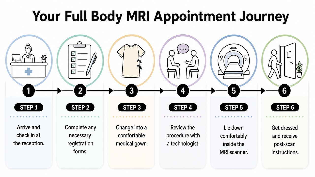
Arrival and check-in
When you arrive, the front desk team confirms your identity and finalizes any remaining forms. If you've already completed much of your paperwork, this part is brief. If not, you'll review medical history, previous surgeries, implants, medications, and any symptoms or concerns that could affect interpretation.
This is also the right time to mention claustrophobia, prior MRI difficulties, back pain, neck pain, or trouble lying flat. Patients sometimes hesitate because they don't want to seem anxious. In reality, the imaging team would rather know early so they can help you prepare.
Changing and final safety review
After check-in, you'll be taken to a private changing area. Depending on the center and what you're wearing, you may change into a gown or MRI-safe clothing. Your belongings are stored securely, and any metallic items are removed before you enter the scanner room.
A technologist then performs a final safety review. This is an intentional pause, not a formality. The team confirms your history, checks for any overlooked metal, and explains how the appointment will unfold.
Entering the MRI suite
The MRI room is typically cool, clean, and uncluttered. The scanner itself looks like a large cylindrical tunnel with a padded table extending from it. Many people expect something harsher or more intimidating. In practice, it usually feels highly structured and purpose-built.
The technologist positions you on the table with pillows, supports, and careful alignment. Comfort matters because staying still is one of the most important parts of a successful scan. You may receive headphones or ear protection, and you'll be shown how to communicate during the exam.
The most reassuring part of the setup is often this. You are not left alone in an uncontrolled situation. The technologist can see you, hear you, and speak with you throughout the study.
During the imaging sequence
Once you're positioned, the table moves gradually into the scanner. The movement is smooth and controlled. You won't feel the images being taken, but you will hear the machine at work.
A full-body MRI is a radiation-free, non-invasive diagnostic tool that uses electromagnetic fields to create detailed images, and for screening it's often completed in under one hour without contrast. In practical terms, that means your role is simple but important. Lie still, listen for instructions, and let the machine do the rest.
Finishing the visit
When the imaging is complete, the table slides out and the technologist helps you sit up slowly. You'll return to the changing area, get dressed, and receive any immediate next-step instructions. Because the scan is non-invasive, you can typically resume the day normally afterward.
Here's the flow in plain terms:
Check in and confirm your information
Change and remove any metallic items
Review safety details with the technologist
Get positioned comfortably on the scanner table
Complete the imaging sequences
Get dressed and leave with guidance on results
That's the full rhythm of the visit. Structured, quiet, and usually much easier than patients imagine beforehand.
What You Will See Hear and Feel Inside the Scanner
The sensory part of MRI is where anticipation tends to build. Once patients know what the sounds and sensations are like, anxiety often drops.
What you'll see
Your visual experience is usually simple. You'll be lying on your back, looking upward or slightly forward depending on your positioning. The inside of the scanner isn't ornate or dramatic. It's a close, rounded medical space with lighting and airflow.
Some people are surprised that the machine itself doesn't feel active in a tactile sense. There's no shaking of the body and no painful sensation from the imaging. The environment feels still, even while the scanner is working intensely around you.
What you'll hear
The sound is the most memorable part for many patients. MRI produces repeated pulses of knocking, tapping, thumping, and buzzing. The patterns can feel mechanical, almost rhythmic. Some sequences sound like distant construction. Others feel more like an electronic beat that starts, stops, and changes tempo.
Headphones or ear protection make a meaningful difference. They won't erase the sound completely, but they soften it and make the experience more manageable. The technologist will also use the communication system to tell you when a sequence is beginning or when a breath-hold is needed.
A short visual overview can help make the setting feel more familiar before you arrive.
What you'll feel
Physically, the main task is stillness. That sounds easy until you're asked to hold that stillness for a sustained period in a confined space. Small adjustments are possible between sequences, but during active imaging the cleaner the stillness, the better the pictures.
If a breath-hold is requested, the technologist will guide you clearly. If no contrast is planned, there's no injection to anticipate. If contrast is ever recommended in a different clinical context, the team should explain why it's being used and what sensation to expect.
For patients comparing providers, this discussion of Prenuvo vs. Longevity Medical Institute in full body MRI may help clarify how protocol and interpretation can shape the experience.
A useful mental shift is to treat the scanner like an audio-rich photography session. The machine is loud because it's working through sequences. Your job is to stay steady long enough for it to gather clean information.
Navigating Claustrophobia and Incidental Findings
Two concerns come up more than any others. The first is claustrophobia. The second is the possibility of finding something unexpected that turns into days or weeks of uncertainty.
Managing claustrophobia in real time
If enclosed spaces make you uneasy, tell the imaging team before the scan begins. That single conversation often changes the experience. Patients usually do better when they know they can communicate at any time, ask for a pause if needed, and use a steady breathing rhythm before the table moves into position.
A few strategies are simple and effective:
Keep your focus narrow. Instead of thinking about the entire scan, think only about the next set of instructions.
Use paced breathing. Slow exhales often help more than forcing deep inhales.
Close your eyes early. Many patients feel calmer if they don't watch the scanner approach around them.
Anchor to the technologist's voice. Familiar cues reduce the feeling of isolation.
If you're prone to claustrophobia, the goal isn't to prove toughness. It's to create a scanning plan that you can actually complete comfortably.
How to think about uncertainty without spiraling
It helps to separate three ideas that often get blended together:
| Situation | What it usually means |
|---|---|
| A finding is detected | The scan noticed a feature |
| A finding is indeterminate | The feature is not fully characterized yet |
| A finding is dangerous | This cannot be assumed without proper review |
Most MRI anxiety doesn't come from the machine itself. It comes from ambiguity after the machine has done its job. That's why pre-scan counseling and careful post-scan interpretation matter so much. If you understand in advance that some findings are common and many prove benign, you're less likely to experience every callback as a crisis.
The LMI Difference AI for Precision and Peace of Mind
The central problem in preventive imaging isn't only detection. It's discrimination. Can the protocol and the reading process separate meaningful abnormalities from harmless biological noise in a way that helps the patient rather than overwhelming them?
That's where advanced imaging design becomes relevant.
Precision begins with protocol
One clinically important protocol uses advanced diffusion-weighted imaging with high b-values of 800 to 1,000 s/mm² and slice thickness of 5 to 7 mm, enabling detection of abnormalities as small as 4 mm, which exceeds a typical 10 mm industry benchmark for cancer sensitivity, as described in this whole-body MRI protocol review in PubMed Central.
Those numbers matter because they reflect how the scan is built, not just how it is marketed. A full body MRI is not one monolithic product. Sequence selection, spatial resolution, and reading workflow all influence whether the final report feels clinically useful.
Where AI fits in
AI should be understood as an assistive layer, not a replacement for radiology. In this setting, it can help flag patterns, organize large imaging datasets, and support consistency across an exam that covers multiple body regions. Used responsibly, that kind of assistance can make review more disciplined and more focused.
At Longevity Medical Institute's AI-enhanced full-body MRI, the stated aim is to combine imaging data with physician context rather than treating every tiny anomaly as equally alarming. That approach is sensible. It doesn't promise perfect certainty, because no screening tool can. It aims for something more realistic and more valuable: better signal, less noise, and clearer next-step decisions.
Better technology is most helpful when it reduces confusion, not when it simply generates more findings.
What peace of mind actually means
Peace of mind doesn't come from a long report filled with technical language. It comes from a report that distinguishes what matters now, what can be monitored, and what is probably incidental. That distinction requires both image quality and clinical judgment.
When readers ask what to expect during a full body MRI, they often mean the scan itself. A more useful question is this. What kind of interpretive system stands behind the scan once the pictures are complete? That is where reassurance becomes either superficial or earned.
Interpreting Your Results and Planning Your Next Steps
After the scan, most patients want the same thing. Not just a report, but a calm explanation of what the report means in real life.
Results should be reviewed in context. That means your images are not interpreted as isolated anatomy alone, but alongside your history, symptoms, family background, and prior testing when available. That clinical layer matters because a finding without context can sound more concerning than it is.
What the follow-up conversation should accomplish
A useful post-scan review does three things well:
Clarifies significance. It separates normal variation, likely benign findings, and issues that warrant further evaluation.
Explains the reasoning. Patients deserve to understand why a recommendation is being made, not just receive a list of next steps.
Creates a plan. If follow-up is needed, the path should be specific and measured rather than vague and alarming.
The psychological and financial toll of incidental findings is often under-discussed. Some studies show 90 to 95% of scans detect some abnormality, most benign, which can create stress and unnecessary biopsy costs, as noted by The Ohio State University Wexner Medical Center's discussion of full-body MRI. That's one reason a thoughtful follow-up matters so much.
How to leave the process with clarity
When you review your report, ask practical questions. Is this finding likely important now, or only worth noting? Does it need another image, a specialist opinion, or only observation? What changes, if any, should you make because of it?
For readers seeking MRI-based diagnostics within a broader preventive framework, MRI diagnostics and regenerative medicine in Los Cabos offers a useful reference point for how imaging can fit into longitudinal care.
A full body MRI is most valuable when it leads to proportionate action. Not panic. Not false reassurance. Just a better-informed next decision.
If you're considering a full body MRI and want guidance on whether it fits your goals, Longevity Medical Institute offers physician-led consultations that place imaging within a broader preventive and longevity framework. Thoughtful screening begins with context, and the right next step is the one that matches your history, risk profile, and priorities.
Author
Dr. Kirk Sanford, DC, Founder & CEO, Longevity Medical Institute. Dr. Sanford focuses on patient education in regenerative and longevity medicine, translating complex therapies into clear, practical guidance for patients.
Medical Review
Dr. Félix Porras, MD, Medical Director, Longevity Medical Institute. Dr. Porras provides clinical oversight and medical review to help ensure accuracy, safety context, and alignment with current standards of care.
Last Reviewed: June 30, 2026
Short Disclaimer
This information is for educational purposes only and is not medical advice. It does not replace an evaluation by a qualified healthcare professional. For personalized guidance, please schedule a consultation.
Published at Longevity Medical Institute Treatments & Resources
Quit referring to autologous stem cells, we only use allogeneic. We produce five different types of stem cells in our biotechnology lab, including placental, Wharton's jelly, adipose, endometrial, and dental pulp.