How Does an AI Full Body MRI Work
It's often assumed that advanced imaging matters only after symptoms start. But what if the more valuable question is this: Can we understand what's happening inside the body before pain, fatigue, or obvious disease forces action?
That question sits at the center of modern longevity medicine. It's also why so many patients now ask, how does an AI full body MRI work, and whether it represents real medical progress or just polished marketing.
The answer is more nuanced than the hype. An AI-enhanced full-body MRI isn't just a “cancer scan.” In the right setting, it's an advanced diagnostic tool that helps physicians investigate anatomy, tissue quality, organ health, and subtle patterns that might otherwise be difficult to appreciate on conventional imaging. Used thoughtfully, it can help build a clearer baseline, support root-cause analysis, and guide more personalized next steps.
That physician-led context matters. Major medical guidelines don't support full-body MRI as a blanket screening test for every healthy adult. But that doesn't mean the technology lacks value. It means the value comes from appropriate use, careful interpretation, and integration into a broader medical strategy.
A New Era of Proactive Health
For decades, medicine has been organized around a familiar sequence. A symptom appears. Testing follows. Treatment begins after a problem becomes obvious enough to name.
That model saves lives, but it's incomplete. Many important changes in the body begin subtly. Tissue can shift, blood vessels can narrow, organ function can change, and structural abnormalities can develop long before someone feels unwell. By the time symptoms arrive, the body may already be compensating for months or years.
That's why proactive diagnostics have become so compelling. Instead of waiting for the body to send an alarm signal, physicians can look for patterns earlier and make decisions from a stronger informational starting point. In that setting, an AI-enhanced full-body MRI becomes less about fear and more about clarity.
A useful way to think about it is this. A standard checkup tells you how the body is performing from the outside. Advanced imaging can show what the body looks like on the inside.
Why this matters in longevity care
In a physician-led longevity program, imaging isn't interpreted in isolation. It's weighed alongside symptoms, family history, lab findings, physical performance, cardiovascular status, and the patient's goals. That's what turns a scan from a standalone event into a clinical tool.
A scan becomes far more useful when it answers a real medical question.
That distinction is central to understanding AI MRI. The technology can make image acquisition and review more advanced, but its ultimate value comes from what a physician does with the information afterward.
Patients who want a deeper look at this broader framework can explore whole-body MRI for longevity and integrated personalized medical care.
From reactive care to informed action
Seen this way, AI full-body MRI belongs to a new category of care. It helps physicians move beyond “Do we see disease?” toward questions like:
What's the current baseline?
Are there subtle structural changes worth watching?
Do imaging findings fit the patient's symptoms or risks?
Should we adjust lifestyle, follow-up testing, or treatment planning?
That's a very different conversation from selling imaging as a universal scan for everyone. It's more precise, more responsible, and ultimately more useful.
Understanding MRI The Foundation of Advanced Imaging
Before AI enters the picture, it helps to understand what an MRI is already doing on its own.
MRI stands for magnetic resonance imaging. It creates detailed pictures of soft tissues throughout the body without using ionizing radiation. That alone is one reason many patients find it appealing, especially when compared with imaging methods that rely on X-rays or radioactive tracers.
What the scanner is actually doing
A simple analogy helps. Think of hydrogen atoms in your body like countless tiny compasses. Under normal conditions, they point in different directions. When you lie inside an MRI scanner, a strong magnetic field lines them up. Radio waves then briefly disturb that alignment, and as the atoms return to their resting state, they release signals.
The scanner captures those signals and turns them into images.
According to the University of Chicago's discussion of the technology, full-body MRI scans align hydrogen nuclei within the body's water molecules using a strong magnetic field and radio waves, generating a 3D map of soft tissues without exposing the patient to ionizing radiation, unlike X-rays, CT, or PET scans.
That's the elegant part. MRI isn't “looking” inside the body the way a camera does. It's measuring how tissues respond to magnetism and radiofrequency energy, then converting that information into detailed images.
Why MRI is so good at soft tissue detail
Different tissues contain different amounts of water and behave differently when the magnetic field and radio waves interact with them. That gives MRI its strength. It can distinguish among organs, muscles, nerves, discs, blood vessels, fat, and many other soft-tissue structures with impressive detail.
If you've ever wondered why MRI is often chosen for the brain, spine, joints, and internal organs, this is why.
A useful comparison:
| Imaging method | What it's especially useful for | Radiation exposure |
|---|---|---|
| X-ray | Dense structures like bones | Yes |
| CT | Fast anatomical overview, trauma, some organ imaging | Yes |
| PET | Metabolic activity | Yes |
| MRI | Soft tissues and detailed structural imaging | No ionizing radiation |
Where patients often get confused
Many people hear “radiology” and assume all imaging uses radiation in the same way. MRI doesn't. It uses magnetism and radio waves, not ionizing radiation.
Practical rule: MRI is about magnetic fields and signal behavior, not the kind of radiation people usually associate with X-rays or CT scans.
That makes MRI particularly attractive when physicians want a thorough look at soft tissues and may need to repeat imaging over time.
How Artificial Intelligence Enhances MRI Scans
Once you understand MRI, the next question becomes easier to answer. How does an AI full body MRI work differently from a conventional MRI?
The short answer is that AI doesn't replace the scanner. It improves what the scanner and radiologist can do together.
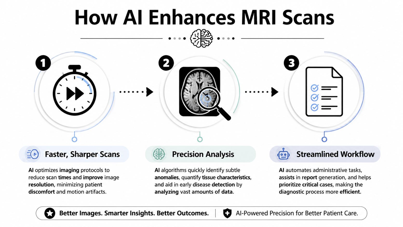
AI as the time-saver
Traditional MRI can be slow because the machine gathers a large amount of data step by step. AI-enhanced systems use deep-learning reconstruction to work with less raw data and still rebuild a high-quality image.
A peer-reviewed review explains that an AI-enhanced full-body MRI uses deep-learning algorithms to reconstruct high-resolution magnetic resonance images from undersampled data, reducing scan time to under 40 minutes while maintaining a signal-to-noise ratio and contrast-to-noise ratio equivalent to conventional scans.
In plain language, “undersampled data” means the scan can collect less information in the usual way, and the AI helps reconstruct what's needed to create a clinically useful image. It's similar to hearing part of a familiar melody and still recognizing the full song because the missing pieces follow patterns the system has learned.
For patients, that can mean less time in the scanner and less chance that movement will blur the images.
AI as the image enhancer
Speed alone wouldn't matter if image quality dropped. That's where the second job comes in.
AI can help denoise MRI images and improve visual clarity. In imaging, “noise” is the graininess or visual interference that can make subtle findings harder to see. When AI improves signal-to-noise and contrast-to-noise ratios, the final images can look cleaner and more defined.
Tiny abnormalities often go unnoticed. A small lesion, a subtle structural change, or a faint difference between healthy and abnormal tissue may be easy to miss if the image is noisy.
Here's a brief visual explanation of the process in motion.
AI as the radiologist's assistant
The third role is often the most misunderstood. AI does not make the final diagnosis. It assists with tasks that are repetitive, time-sensitive, and highly quantitative.
Based on verified data about FDA-cleared systems such as Ezra Flash and Ezra Assist, AI can help with:
Reconstruction of images: Turning limited acquisition data into clinically useful high-resolution imaging.
Measurements and annotations: Assisting radiologists with structured review tasks.
Segmentations: Separating organs or regions so the anatomy can be evaluated more precisely.
Detection support: Making subtle pathologies easier for the radiologist to appreciate.
One verified source describes FDA-cleared AI tools such as Ezra Flash and Ezra Assist as improving image quality by reducing noise and improving signal-to-noise and contrast-to-noise ratios, helping radiologists detect small pathologies such as early-stage tumors, small brain aneurysms, and signs of neurodegeneration, as described in this overview of MRI with AI as a preventative diagnostics tool.
The best analogy
If conventional MRI is like taking a detailed photograph in dim light, AI helps in three ways:
It lets the camera work faster.
It sharpens the image afterward.
It gives the expert reviewer better tools for measuring what's in the frame.
AI in MRI is best understood as an advanced assistant. It helps the human expert see more clearly and work more efficiently, but it doesn't replace clinical judgment.
That combination is what makes the technology important.
The Clinical Advantages of an AI Full Body MRI
The patient benefits become clearer once you separate technical novelty from clinical function. AI-enhanced MRI isn't useful because it sounds advanced. It's useful when it improves the actual scan experience and the quality of information a physician can act on.
What patients notice first
The first advantage is practical. A shorter scan is usually easier to tolerate.
That matters more than many people realize. Long scans can be uncomfortable for patients with back pain, claustrophobia, stiffness, or difficulty remaining still. A more efficient process can reduce motion artifacts and improve image quality because the patient can hold a comfortable position more consistently.
What physicians value most
The deeper advantage is better interpretability. When AI improves image clarity and supports measurements or segmentation, radiologists can review anatomy with greater precision. That can strengthen confidence when assessing subtle findings and help create more useful comparisons over time.
This is especially relevant when a physician is not just asking, “Is something obviously wrong?” but also, “Do these findings help explain the patient's symptoms or long-term risk?”
Conventional MRI vs AI-Enhanced MRI
| Feature | Conventional Full Body MRI | AI-Enhanced Full Body MRI |
|---|---|---|
| Scan duration | Typically longer | Can be significantly shorter |
| Image reconstruction | Traditional reconstruction methods | Deep-learning reconstruction from undersampled data |
| Image noise | More dependent on conventional acquisition limits | AI can reduce noise and improve clarity |
| Workflow | More manual review tasks | AI can support measurements, annotations, and segmentation |
| Patient experience | Longer time lying still | More efficient and often easier to tolerate |
Patients considering physician-guided imaging can learn more about MRI diagnostics in regenerative medicine in Los Cabos.
Why these differences matter clinically
AI-enhanced MRI can support earlier appreciation of subtle structural changes, provide more consistent quantitative review, and help organize large imaging datasets in a clinically useful way. It may also support ongoing tracking when a physician wants to monitor an area over time.
That doesn't mean every scan reveals something important. It means the scan has become a more refined instrument when there is a legitimate reason to look.
Your Scan at The Longevity Medical Institute
For many patients, the technology feels less intimidating once they understand the actual experience. The process is structured, physician-led, and designed to turn raw imaging data into a useful medical conversation.
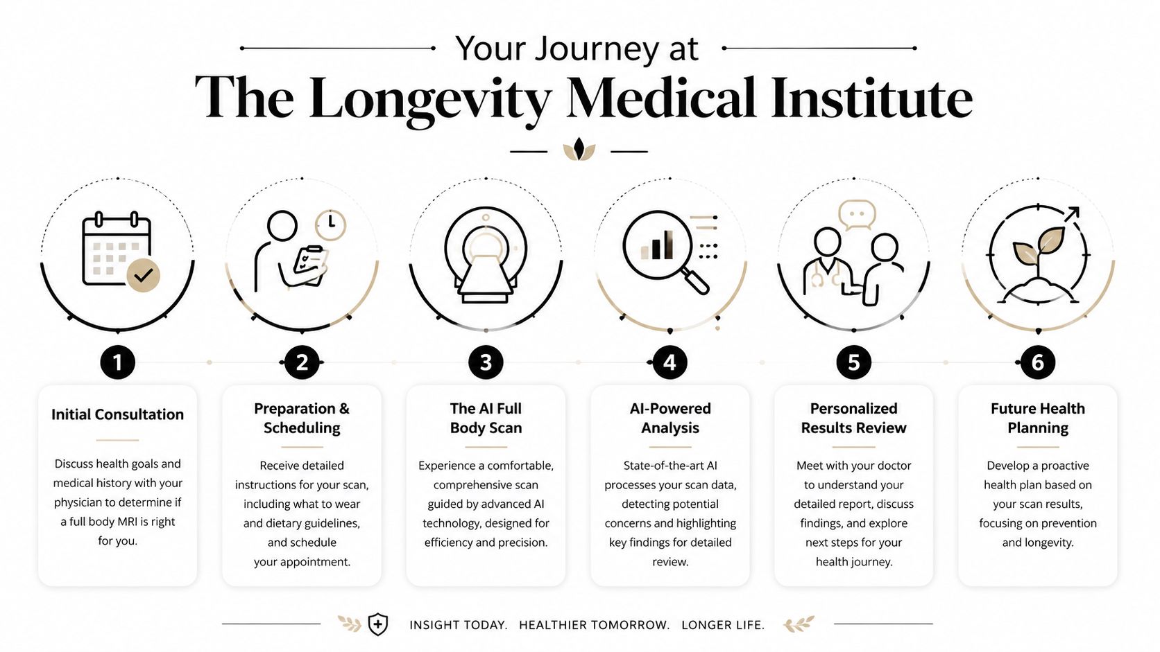
It begins with the right question
A full-body MRI should start with medical context, not impulse. Before imaging, the physician reviews symptoms, history, prior testing, family patterns, and the patient's goals. That determines whether broad imaging is likely to add clarity or create noise.
A physician-led program differs from a consumer scan model. The purpose is not to generate a long list of incidental findings. The purpose is to investigate meaningful patterns and build a more informed strategy.
Patients comparing different approaches often ask thoughtful questions about protocol, interpretation, and follow-up. That's why many review common questions about Prenuvo versus a physician-led longevity MRI approach.
What the day of the scan feels like
During the scan, you'll lie still while the MRI system captures images across the body. The environment is designed around comfort and efficiency, since patient movement affects image quality. The scan itself remains noninvasive.
After acquisition, AI-assisted processing helps organize and refine the imaging data. But the important part comes next. A radiologist reviews the images, and the findings are interpreted in the context of the patient's medical picture rather than handed over as isolated observations.
What happens after the report
The value of imaging often shows up after the scan, not during it.
At Longevity Medical Institute, MRI findings can be integrated with physician assessment, broader diagnostic workup, and personalized planning. That may include follow-up imaging, laboratory correlation, cardiovascular evaluation, movement and musculoskeletal review, or a regenerative medicine discussion when clinically appropriate.
A sophisticated scan is only the beginning. The real work is translating anatomy into decisions.
That translation is what patients tend to remember. They don't just leave with images. They leave with interpretation, prioritization, and a clearer sense of what matters now versus what needs observation.
Safety Limitations and the Importance of Clinical Guidance
The most trustworthy explanation of AI full-body MRI includes its limitations.
This technology can be powerful, but it isn't a universal screening solution for everyone. In fact, one of the biggest mistakes in this space is assuming that a more advanced scan automatically means a better test for any person in any situation.
Where the scan is most useful
AI-enhanced full-body MRI tends to make the most sense when physicians are:
Investigating symptoms: Looking for structural explanations when standard evaluation hasn't fully answered the question.
Clarifying risk: Especially when family history, prior disease, or other factors raise concern.
Establishing a baseline for longevity: Being proactive by creating a detailed internal map that can be tracked over time.
Integrating diagnostics: Using imaging alongside labs, examination, and specialist review.
Patients who want to understand how a responsible clinic approaches these issues can review safety and transparency in advanced medical care.
Why physician oversight matters
AI can accelerate scans. It can sharpen images. It can help with segmentation and measurements. What it cannot do is decide what a finding means for your life, your risk, or your next step.
The more detailed a scan becomes, the more important clinical judgment becomes.
That's why physician guidance isn't an optional extra. It's the safeguard that turns complex data into careful medicine.
A New Foundation for Personalized Medicine
The most meaningful way to think about AI-enhanced MRI is not as a one-time event, but as a foundation layer of personalized medicine.
A detailed full-body scan can provide a broad anatomical baseline. When that information is interpreted alongside symptoms, labs, cardiovascular evaluation, and ongoing follow-up, physicians can make decisions with a level of specificity that guesswork can't provide.
The technology moves beyond the narrow idea of “finding disease.” It can help frame organ health, structural patterns, musculoskeletal balance, and other measurable features in a way that supports a long-term plan rather than a single moment of detection.
According to the Institute's overview of this approach, the AI-driven analytics layer supports identification of hundreds of wellness indicators, including organ health and musculoskeletal balance, offering actionable data for personalized regenerative medicine plans.
What that changes for patients
When imaging is integrated well, patients and physicians can make more focused choices. They can decide what deserves immediate action, what should be monitored, and where supportive interventions may have the greatest value.
That's one reason digital continuity matters too. Many patients now expect their imaging, lab results, and care plans to live inside one coherent system, not across disconnected reports. A useful example is the Longevity Patient App and its 360-degree view of health data.
The promise of AI MRI isn't that it replaces medicine. It's that it gives medicine a better map.
Author & Medical Review Information
Author
Dr. Kirk Sanford, DC, Founder & CEO, Longevity Medical Institute. Dr. Sanford focuses on patient education in regenerative and longevity medicine, translating complex therapies into clear, practical guidance for patients.
Medical Review
Dr. Félix Porras, MD, Medical Director, Longevity Medical Institute. Dr. Porras provides clinical oversight and medical review to help ensure accuracy, safety context, and alignment with current standards of care.
Last Reviewed: June 28, 2026
Short Disclaimer
This information is for educational purposes only and is not medical advice. It does not replace an evaluation by a qualified healthcare professional. For personalized guidance, please schedule a consultation.
We produce five different types of allogeneic stem cells in our biotechnology lab, including placental, Wharton's jelly, adipose, endometrial, and dental pulp.
If you'd like to explore physician-led diagnostics and longevity care, visit Longevity Medical Institute and browse articles published under Treatments & Resources to learn more about AI-integrated imaging, regenerative medicine, and personalized health planning.