

Comprehensive Heart Assessment: The Complete Patient Guide
Only 1 in 5 people in the U.S. had optimal heart health in the American Heart Association's 2022 analysis of Life's Essential 8, while about 80% fell into low to moderate cardiovascular health, a reminder that heart disease usually develops over time long before it becomes urgent AHA Life's Essential 8 analysis. That's why a Heart Assessment matters. It doesn't wait for chest pain, shortness of breath, or an abnormal stress test to start paying attention, it looks for the biology, the arteries, the muscle, the structure, and the electrical system that can fail long before symptoms arrive.
Traditional screening can be useful, but it often samples only one slice of risk. A blood pressure check tells you about pressure in the moment, a cholesterol panel gives you part of the metabolic picture, and a resting ECG shows one snapshot of electrical activity. None of those, by itself, gives the full story. A real preventive evaluation layers them together, then adds imaging and biomarkers so a clinician can see how the heart is behaving from multiple angles, much like comparing a map, a traffic report, a building inspection, and a wiring diagram before making a major decision.
The goal is simple. Thorough should mean more than “more tests.” It should mean a more complete way to understand where risk is hiding and what to do about it before disease becomes irreversible.
Why Heart Disease Still Wins and What a Thorough Heart Assessment Changes
Heart disease stays dangerous because it usually develops without obvious signs. Plaque can accumulate in the coronary arteries, the heart muscle can weaken or scar, valves can leak, and rhythm problems can develop while the person still feels mostly fine until a major event forces attention. That silent phase is where prevention has the most value, and also where partial testing fails most often.
The American Heart Association's Life's Essential 8 framework makes that wider problem easier to see. It measures eight modifiable domains, diet, physical activity, nicotine exposure, sleep, body mass index, blood lipids, blood glucose, and blood pressure, which is a much better reminder that cardiovascular health has multiple moving parts than a single number ever could. The practical message is simple, a meaningful assessment has to look beyond one blood pressure reading or one cholesterol value and review several risk factors together.
The five dimensions that organize a real evaluation
A useful way to think about cardiovascular care is to separate the heart into five dimensions. Biological risk covers the internal signals that predispose someone to disease, including inflammation and metabolic stress. Coronary artery health asks whether the vessels feeding the heart are already developing plaque. Heart muscle health looks at the myocardium itself, the part that pumps.
Two more dimensions matter just as much. Structural and functional health covers the chambers, valves, and blood flow mechanics. Electrical health covers rhythm and conduction, the system that tells the heart when to beat and how regularly.
Practical rule: if a test only looks at one dimension, it can still be useful, but it cannot be called complete.
That is why the standard question is not “What test should I get?” It is “Which dimensions of risk matter most for me, and which tools can see them?”
For readers comparing options in actual practice, it helps to distinguish a thorough preventive program from general education resources like preventing heart disease, because many public-facing guides stop at general lifestyle advice and never reach the level of multimodality evaluation. A broader clinical framework starts with the assumption that no single exam can reveal everything that matters, and the same logic underlies cardiology and longevity care.
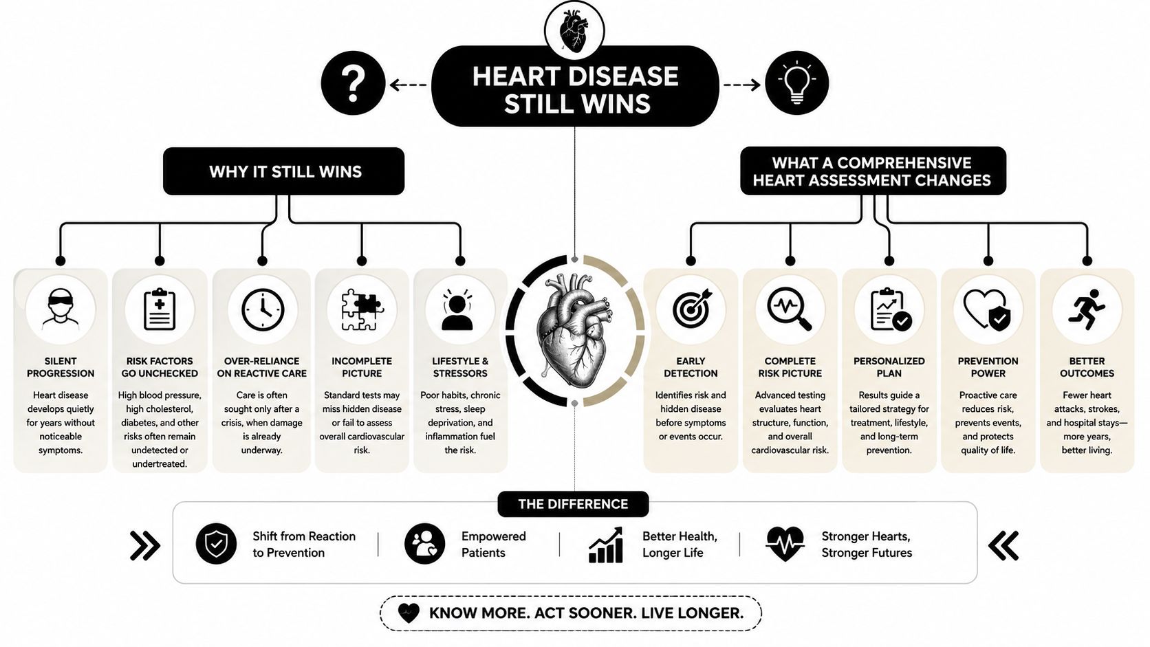
The shift is philosophical as much as technical. A reactive model waits for an event. A preventive model looks for pattern changes first, then uses the right tool for each layer of risk.
The Clinical Foundation, History, Physical Exam, ECG, and Advanced Cardiovascular Biomarkers
A careful cardiac assessment still starts with the basics, because the first clues often appear in the story, not the scanner. The history should ask about chest-pain quality, whether symptoms happen with exertion or at rest, how far they radiate, and whether there is a family history of sudden death or inherited cardiac conditions. Those patterns help sort risk into likely coronary disease, rhythm problems, structural disease, or inherited syndromes.
What a careful exam actually looks for
A focused cardiovascular exam is more than listening over the chest. It includes inspection, palpation, auscultation, peripheral pulses, blood pressure in both arms, jugular venous distension, edema, thrills, and the point of maximal impulse in the fifth intercostal space at the midclavicular line. Carotid pulses are auscultated for bruits before palpation to reduce stroke risk, a good reminder that technique matters as much as interpretation Merck Manual cardiovascular examination.
A standard focused exam also moves in a deliberate order, general appearance, skin, nails, mouth, neck, extremities, pulses, veins, chest, heart sounds, lungs, and the abdomen or lower limbs when indicated NCBI focused cardiovascular exam. That sequence helps clinicians avoid missing peripheral signs of low perfusion or systemic disease.
The listening step deserves special attention. Teaching materials describe five auscultation areas, and some use a classic six-point sequence across the aortic, pulmonic, tricuspid, and mitral areas. Clinicians should identify S1 and S2 and document murmurs by timing, location, radiation, and intensity. Peripheral circulation belongs in the same examination, with bilateral radial, brachial, dorsalis pedis, and posterior tibialis pulses, plus capillary refill documented as less than or greater than 2 seconds focused assessment manual.
The ECG sets the electrical baseline
A 12-lead ECG gives a fast, noninvasive view of rhythm and conduction. It can reveal atrial fibrillation, arrhythmias, ischemic changes, chamber enlargement, and evidence of prior silent injury. In a full cardiac program, the ECG is the electrical map that can be compared over time, the same way a clinician would compare two drawings of the same circuit to see where the wiring has changed.
Advanced biomarkers deepen the biological readout
Biomarkers make prevention more precise because they can show inherited risk, inflammation, metabolic dysfunction, and wall stress before symptoms appear.
| Advanced Cardiovascular Biomarker | What It Reveals |
|---|---|
| Lipoprotein(a) | Inherited lipid risk that can persist despite otherwise acceptable standard lipids |
| ApoB | The number of atherogenic particles that can drive plaque formation |
| ApoA1 | A key component of HDL-related biology and reverse transport pathways |
| ApoB/ApoA1 ratio | The balance between atherogenic and protective lipoprotein patterns |
| hs-CRP | Inflammatory activity that can amplify vascular risk |
| NT-proBNP | Cardiac wall stress and strain |
| Homocysteine | A metabolic marker that can reflect broader vascular risk biology |
| Insulin | Early metabolic strain and compensatory hyperinsulinemia |
| HOMA-IR | A practical estimate of insulin resistance |
| Hemoglobin A1c | Longer-term glucose exposure and glycemic burden |
A standard lipid panel can miss inherited or particle-based risk, and it does not show whether inflammation or insulin resistance is already pushing the system in the wrong direction. A thorough heart assessment reads these signals together, then uses them to decide whether imaging, medication, or lifestyle changes deserve priority.
For readers who want a structured look at this lab layer, the resource on advanced cardiovascular biomarkers is a useful complement to the clinical discussion here.
Coronary Artery Health, 128-Slice Coronary CT, Calcium Scoring, and CT Angiography
Coronary artery disease is the most common cause of heart attacks, and it can remain undetected for years while plaque accumulates in the vessel wall. That's why imaging the arteries is so important. You can't see coronary plaque with a stethoscope, and a normal resting ECG doesn't rule it out.
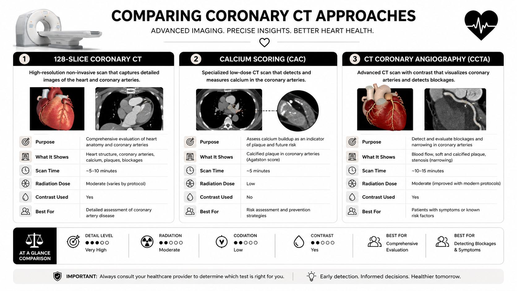
A 128-slice coronary CT represents next-level imaging because it acquires data quickly and with higher resolution, which improves visualization of coronary anatomy and helps clinicians detect plaque and narrowing more accurately. In practical terms, it's closer to a high-definition plumbing inspection than a simple snapshot. The distinction matters because the coronary arteries are the pipes that feed the heart muscle, and if they're developing disease, the rest of the system is at risk.
Calcium scoring versus CT angiography
These two tests answer different questions.
Coronary artery calcium scoring is a fast, low-dose study that measures calcified plaque burden. It doesn't require contrast, and it's useful when the main question is how much calcified atherosclerosis is present. Coronary CT angiography, by contrast, uses contrast and visualizes both calcified and soft plaque while also showing the degree of narrowing in the arteries.
| Test | Main Use | Strength | Limitation |
|---|---|---|---|
| Calcium scoring | Estimate calcified plaque burden | Fast, low-dose, no contrast | Doesn't show soft plaque or lumen detail as fully |
| CT angiography | Evaluate plaque and narrowing | Sees calcified and soft plaque, shows anatomy directly | Requires contrast and more preparation |
That difference is why one test doesn't replace the other. A calcium score can be useful for broad risk stratification, while CT angiography is better when a more detailed look at coronary anatomy is needed. A thorough program chooses based on the clinical question, not on the loudness of the imaging label.
Coronary CT evaluates the pipes. It doesn't evaluate the muscle that those pipes feed, and that's why the rest of the assessment still matters.
For readers trying to understand this technology in a broader diagnostic context, the internal overview of AI full-body MRI helps show how coronary CT fits into a larger imaging strategy. The conceptual takeaway is simple, the scan should match the question. If the concern is hidden plaque, CT is the right lens. If the concern is muscle injury or scarring, another modality is needed.
Heart Muscle Health, AI-Enhanced Cardiac MRI for Myocardial Evaluation
If coronary CT looks at the arteries, cardiac MRI looks at the heart muscle itself. That distinction matters in a true heart assessment, because a person can have reassuring arterial findings and still have meaningful myocardial disease. In practice, cardiac MRI is often treated as the gold standard for evaluating the myocardium because it can show function and tissue character in the same study.
AI-enhanced post-processing strengthens that role by making measurements more reproducible and by improving workflow efficiency. The value is not speed alone. It is clarity. A carefully interpreted cardiac MRI can quantify ventricular volumes, ejection fraction, cardiac output, myocardial inflammation, fibrosis, scar tissue, cardiomyopathies, structural heart disease, and prior silent cardiac injury.
Why MRI adds information other tests can't
Echocardiography can assess function well, but MRI gives exceptional tissue detail. CT is excellent for coronary anatomy, but it does not characterize the myocardium in the same way. MRI helps clinicians sort out whether a weakened pump is due to ischemia, inflammation, scarring, infiltrative disease, or a cardiomyopathic process.
That is why CT and MRI work as complements. Coronary CT evaluates the arteries. Cardiac MRI evaluates the muscle. A complete assessment uses both when the clinical question and the risk profile justify it.
Clinical rule: if the question is “Are the vessels narrowed?” CT answers that. If the question is “What happened to the muscle?” MRI is the stronger tool.
Stress imaging adds another layer when the question is flow under demand. Stress cardiac MRI is usually used when clinicians need to diagnose or exclude obstructive coronary disease, or when symptoms and ECG findings raise concern. A resting MRI is often enough when the main question is structure, function, or tissue characterization. That difference matters, because broad screening and symptom-driven diagnostics are not the same task, and advanced imaging should be chosen with the question in mind.
A physician who wants to place MRI into a broader preventive strategy can also look at what an AI full-body MRI is to understand how the modality fits into body-wide risk review. For the heart specifically, the point is sharper. MRI answers questions about the myocardium that no coronary test can answer.
Structural Function and Electrical Health, Thorough Echocardiography and the AI-Integrated 12-Lead ECG
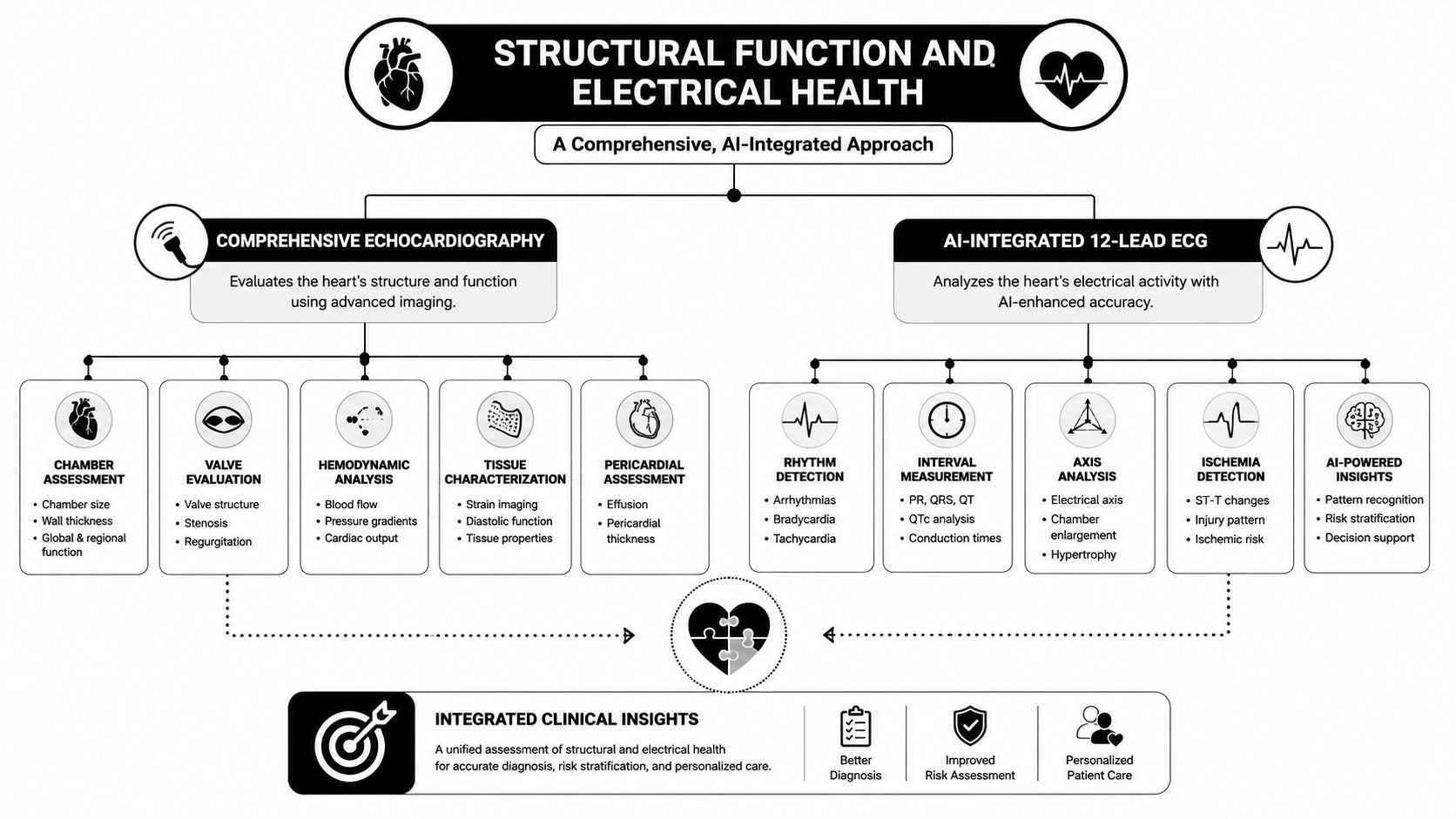
A complete heart assessment has to examine both the pump and the wiring, because one can look acceptable while the other is already showing strain. Echocardiography evaluates the mechanical side, while the 12-lead ECG evaluates the electrical side. They are often described as support tests, but in a preventive cardiology workup they are major pillars of the five-dimension framework, structural health and electrical health.
Echocardiography in real time
A proper transthoracic echocardiogram includes demographic and technical documentation, image-quality assessment, and indexed quantitative measurements of all chambers, along with valve structure and function and diastolic parameters complete transthoracic echocardiogram standards. Standard views include parasternal long- and short-axis and subcostal long- and short-axis sweeps. Those views help detect congenital connections, chamber enlargement, outflow-tract abnormalities, and valvular hemodynamic lesions that can be missed on auscultation alone.
That makes echo more than a “heart ultrasound.” It is a live mechanical audit. It shows how well blood moves, whether the chambers are enlarged, whether valves open and close properly, and whether systolic or diastolic function is impaired. A scan done well can separate a stiff ventricle from a leaky valve, which are very different problems even if the symptoms overlap.
A clearer image also depends on the equipment used to capture it. In many diagnostic settings, a SonoSite Edge II ultrasound platform helps clinicians obtain and review the heart's motion at the bedside, where timing and angle can change what is seen.
The ECG and AI-assisted interpretation
A 12-lead ECG provides the electrical counterpart. It can identify rhythm problems, conduction delays, atrial fibrillation, chamber enlargement, ischemic patterns, and evidence of silent prior injury. AI-assisted analysis can strengthen pattern recognition, especially when the tracing contains subtle abnormalities that are easy to miss, but physician interpretation is still essential. The machine can flag, the clinician decides.
That pairing matters because electrical problems and mechanical problems often influence one another. A person can have a murmur and normal rhythm, or a normal-feeling heartbeat and a concerning ECG. A good assessment checks both and compares them side by side, like looking at both the engine and the dashboard before deciding whether a car is healthy.
The point is not to choose one tool and ignore the other. Echocardiography describes structure and motion, while the ECG records the heart's electrical pattern, and together they create a fuller picture of how the heart is functioning in real life.
Acoustic and AI-Auscultation Layers, CADScor and the Eko CORE 500 With SENSORA
Some heart risk never shows up on imaging first, it shows up as sound. That's why acoustic tools belong in a full-scope program. They add another layer between the stethoscope and the scanner, and that layer can be especially useful when clinicians want a radiation-free, point-of-care look at hidden risk.
CADScor uses AI-powered acoustic analysis to help assess the likelihood of coronary artery disease without radiation or contrast. It's not a replacement for imaging, but it can help triage which patients need deeper evaluation. Eko CORE 500 with SENSORA combines digital auscultation with AI-assisted analysis to help identify murmurs, valve abnormalities, and other findings that can be subtle on a traditional exam.
These tools fit neatly into prevention because they're quick, repeatable, and low burden for the patient. They're also useful when contrast isn't ideal or when a clinician wants extra data before moving to more advanced imaging.
A simple way to think about it is this. Imaging sees structure. Acoustic analysis hears physiology. Together, they can surface patterns that one modality alone might miss.
AI-auscultation works best when it supports, not replaces, a careful clinician exam.
The value isn't that the device is louder or shinier. The value is that it gives the physician more information at the bedside, which can refine the next step in a diagnostic pathway. In a program, that's the entire point, better triage, better targeting, and fewer blind spots.
How the Five Dimensions Come Together at Longevity Medical Institute
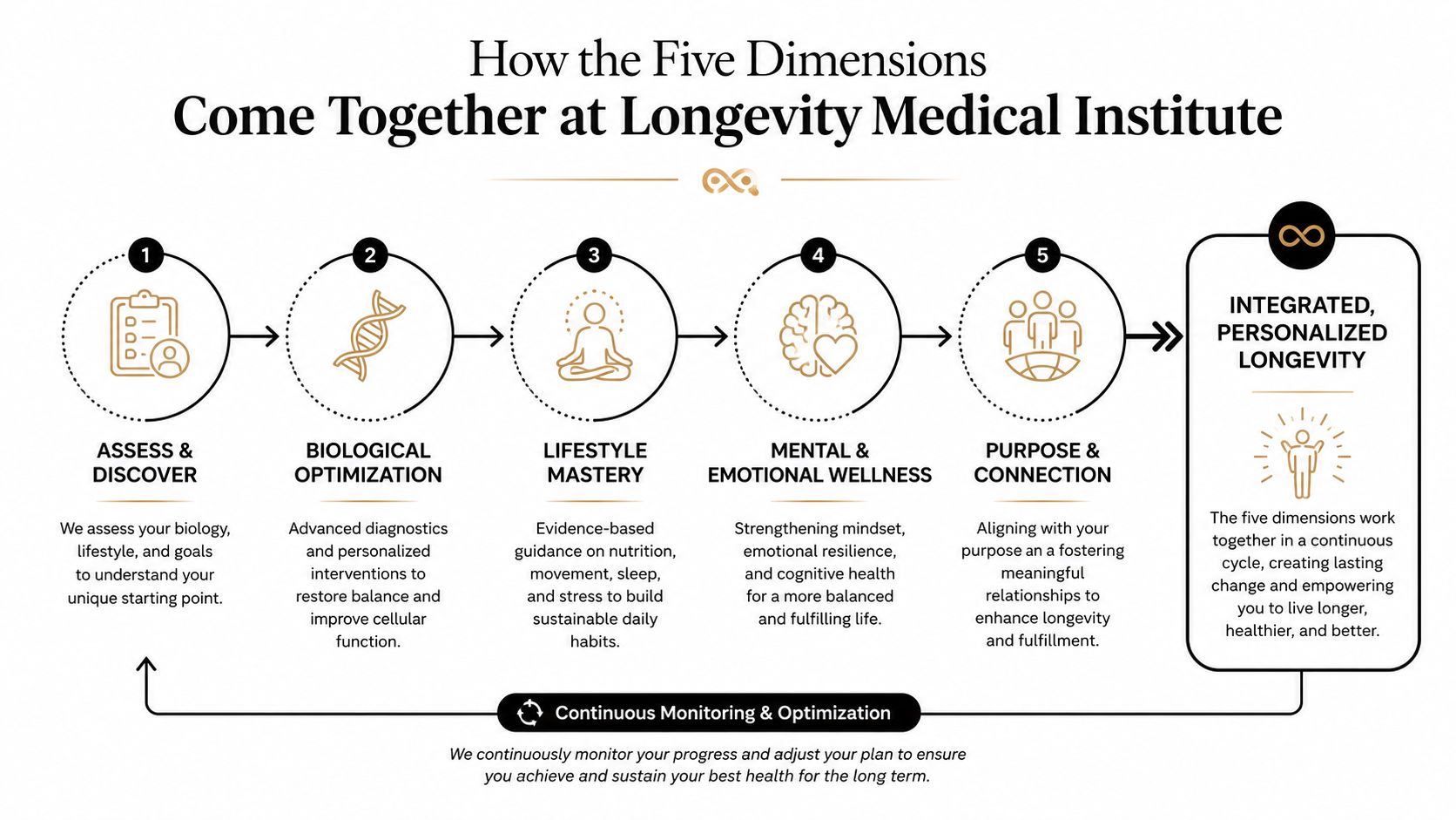
A preventive heart assessment works best when each dimension has a specific tool attached to it. Biological risk is measured with advanced biomarkers from an in-house clinical laboratory, coronary artery health is evaluated with 128-slice coronary CT and calcium scoring or angiography, heart muscle health is assessed with AI-enhanced cardiac MRI, structural and functional health is studied with echocardiography, and electrical health is assessed with the AI-integrated 12-lead ECG. CADScor and the Eko CORE 500 add the acoustic layer that traditional screening often skips.
What the workflow feels like
In practical terms, this kind of evaluation usually starts with preparation. Patients may be asked to fast for certain labs, stay hydrated, review medications, and avoid wearing clothing that interferes with imaging or ECG placement. Contrast decisions matter for coronary CT angiography and some MRI protocols, so the clinical team should review kidney function, allergy history, and any prior contrast reactions before scheduling.
The interpretation stage is where the program becomes more than the sum of its parts. A physician-led review combines the lab findings, the imaging findings, the ECG, the acoustic data, and the physical exam into one cardiovascular risk profile. That allows the clinician to separate reassuring findings from actionable ones instead of treating each result in isolation.
The Longevity Patient App can help organize this kind of multi-source health information in a way that patients can use after the visit. The point is not just to collect data, it's to make the data understandable enough to guide decisions.
What makes the program coherent
The strength of this model is integration. Biomarkers tell you whether biology is moving in the wrong direction. CT tells you whether plaque is present. MRI tells you what the muscle looks like. Echo tells you how the heart moves. ECG tells you how it conducts. CADScor and Eko add acoustic signals that round out the picture.
That's how a heart assessment becomes a risk map rather than a test menu.
Frequently Asked Questions About a Thorough Heart Assessment
Who should consider it
Adults with risk factors, a strong family history, prior abnormal tests, or prevention goals often benefit from a broader assessment. People who feel well but want a serious longevity-focused baseline can also be appropriate candidates, especially when the question is not whether disease is present, but where risk may be hiding.
How often should it be repeated
That depends on the initial findings, the person's risk profile, and what was done at baseline. Someone with reassuring labs and imaging may not need the same cadence as someone with plaque, metabolic risk, or a concerning family history. The interval should be individualized by the physician reviewing the case.
Does the imaging involve radiation
Coronary CT does involve radiation, while coronary calcium scoring generally uses less than contrast-based CT angiography. MRI does not use ionizing radiation. The right study depends on the clinical question, and the goal is always to choose the least invasive tool that still answers the question well.
How is this different from an executive physical
An executive physical often focuses on broad screening. A preventive heart assessment goes further by matching the test to the cardiovascular dimension being evaluated, biology, coronary arteries, myocardium, structure, or electrical function. That difference matters because “normal” on one screen does not necessarily mean low risk across the board.
What happens after the assessment
Findings can lead to reassurance, follow-up imaging, medication changes, nutrition or activity guidance, or referral to another specialist. The value of the assessment is not just detection, it is direction. A good report should leave you with a clear explanation of what is normal, what needs watchful waiting, and what needs action now.
For patients who want a physician-led evaluation that brings these pieces together, the most useful next step is a consultation that can match the right test to the right risk. The most effective treatment for heart disease is still early identification of risk, before symptoms begin.
Author
Dr. Kirk Sanford, DC, Founder & CEO, Longevity Medical Institute. Dr. Sanford focuses on patient education in regenerative and longevity medicine, translating complex therapies into clear, practical guidance for patients.
Medical Review
Dr. Félix Porras, MD, Medical Director, Longevity Medical Institute. Dr. Porras provides clinical oversight and medical review to help ensure accuracy, safety context, and alignment with current standards of care.
Last Reviewed: July 25, 2026
Short Disclaimer
This information is for educational purposes only and is not medical advice. It does not replace an evaluation by a qualified healthcare professional. For personalized guidance, please schedule a consultation.
Published in the treatments and resources library at Longevity Medical Institute Treatments & Resources.
A CTA for Longevity Medical Institute. Schedule a physician-led preventive cardiovascular assessment to review your biological risk, coronary artery health, heart muscle health, structural function, and electrical health in one coordinated workup, then leave with a clear plan aligned to your goals and findings.