

Early Heart Disease Detection: A Precision Guide
You can train hard, eat clean, and feel completely fine, then walk into a concierge cardiac visit expecting reassurance. Sometimes that visit is exactly where early heart disease detection changes the story, because the first clear sign of coronary risk isn't chest pain, it's often a finding that was already there, before symptoms ever appeared.
That gap between feeling well and being well is where modern prevention earns its keep. A layered cardiac evaluation can reveal plaque, electrical abnormalities, inflammation, or vascular dysfunction long before a first event forces the issue, which is why precision screening is becoming the standard for people who want to protect performance, travel with confidence, and make decisions from data rather than guesswork.
When the Heart Signals Nothing Yet Still Speaks
A 47-year-old executive comes in after a routine trip and says he feels great. He lifts weights, takes his recovery seriously, and keeps his annual physicals on schedule. Then advanced cardiac imaging shows silent plaque, and a calcium score reframes his risk from “probably fine” to “needs a prevention plan now.”
That story is common because the heart can tolerate gradual disease for years before it complains. The arteries don't always send pain as a warning, and routine habits can look impressive while plaque continues to form under the surface. In that sense, absence of symptoms is not evidence of absence of disease.
Why silent disease is so easy to miss
People often assume a normal treadmill, a good resting pulse, or a clean feeling after exercise means low risk. Those signals matter, but they don't see everything. Coronary disease can progress until a first event exposes what earlier testing might have found in time.
The CDC estimates that 5.0% of adults had ever been diagnosed with coronary heart disease in 2024 CDC fast facts. The same source notes roughly 580,000 first myocardial infarctions and 610,000 first strokes each year in the United States, which is a reminder that first events still happen often, even in the era of better screening and treatment CDC fast facts.
Practical rule: If the question is “Do I feel fine?”, the better clinical question is “What is my biology doing before I feel anything?”
A premium prevention approach treats those questions differently. One checks comfort. The other checks risk. That distinction is why high-performance patients, frequent travelers, and people building long-term health plans increasingly want more than a routine exam.
Why Early Detection Has Mattered Over Time
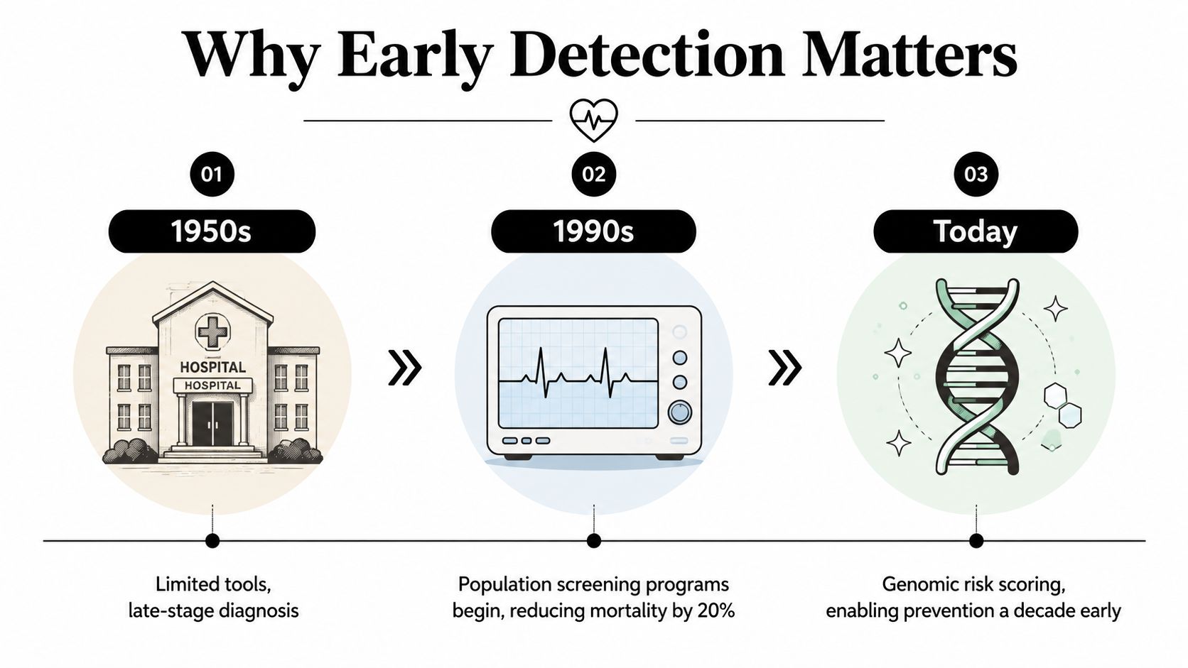
A patient may feel well and still carry years of silent arterial change. That is why early heart disease detection has never been about one dramatic test. It has been about seeing risk sooner, while there is still time to change the course.
The historical record shows why this matters. In the United States, among adults aged 25 to 64, the age-standardized premature heart disease mortality rate fell by 70%, from 206 deaths per 100,000 in 1968 to 62 per 100,000 in 2017 CDC mortality report. Over the same period, the share of all premature deaths attributable to heart disease dropped from 33% in 1968 to 1977 to 19% in 2008 to 2017 CDC mortality report.
That change came from earlier recognition of risk factors, better access to cardiovascular care, and better treatment after disease was detected. The burden did not disappear, though, because there were 126,842 deaths in 2017, or about one death every 4 minutes CDC mortality report.
What that history means for a modern patient
The lesson is straightforward. When clinicians find risk earlier, outcomes improve at the population level, and patients gain more options before symptoms appear. That is the reason prevention programs now focus on upstream detection rather than waiting for a crisis.
For a patient choosing between screening pathways, the structure of the program matters as much as the test itself. A coordinated evaluation that brings together risk review, biomarkers, and imaging is different from a single isolated order, and a resource like Longevity Medical Institute's preventive cardiology overview reflects that layered approach. For patients seeking concierge-level cardiac evaluation, including medical travelers and high-net-worth patients who want a more personalized pathway, the point is simple. Earlier identification gives the clinician more room to act, and more room to act usually means better choices.
Mapping Your Personal Cardiovascular Risk Profile
A useful risk profile starts with the basics, but it doesn't stop there. Blood pressure, lipids, smoking, diabetes, and family history still matter, yet they don't capture every person who is moving toward disease. That's why preventive cardiologists think in layers.
The risk enhancers people often overlook
Some of the most important enhancers sit outside the standard checkbox exam. Lp(a) levels that are higher than normal, higher apoB, pregnancy-related hypertension, premature menopause, tobacco exposure, and chronic inflammation can all change how seriously a clinician interprets a “normal” looking profile. In younger adults, those details matter even more because routine screening can miss early atherosclerosis in the 20s and 30s.
A JACC state-of-the-art review says clinicians should look beyond traditional risk factors and consider nonfasting lipids, Lp(a), apoB, and selected CAC scoring in young adults JACC review. That kind of thinking helps answer the question many patients really have, which is not “Do I have any risk at all?” but “What kind of risk do I have, and how early should I look?”
An organized way to approach this is to ask three questions at the same visit.
What is my baseline risk? Blood pressure, lipids, glucose, family history, smoking status, and age still establish the starting point.
What changes the interpretation? Lp(a), apoB, pregnancy history, premature menopause, inflammatory conditions, and tobacco exposure can shift the conversation.
What level of testing matches my profile? Some patients need only laboratory refinement. Others need imaging or rhythm assessment because their biology is more revealing than their routine exam.
The most useful risk profile is the one that changes what you do next, not the one that just sounds reassuring.
Biomarkers That Reveal What a Routine Lipid Panel Misses
A routine lipid panel tells part of the story. It shows cholesterol levels in broad strokes, but it can miss how many atherogenic particles are circulating, how much inflammatory activity is present, and whether the vascular lining is under stress. That is why precision programs use biomarkers as a second lens, not a replacement for clinical judgment.
Comparing the common tests
| Biomarker | What It Measures | Clinical Question Answered |
|---|---|---|
| Total cholesterol | Overall cholesterol burden | Is the broad cholesterol level elevated? |
| LDL-C | Cholesterol carried by LDL | How much LDL cholesterol is present? |
| ApoB | Number of atherogenic particles | How many plaque-forming particles are circulating? |
| Lp(a) | A genetically influenced lipoprotein particle | Is inherited risk likely being missed? |
| High-sensitivity CRP | Low-grade inflammation | Is inflammatory burden adding to vascular risk? |
| Fasting glucose | Blood sugar level at the time of testing | Is dysglycemia part of the risk picture? |
| Advanced imaging-linked markers | Functional or structural vascular clues | Is disease present before symptoms? |
That table becomes more useful when the goal is risk reclassification. A patient with a modest LDL-C but high apoB can look “normal” on the surface while still carrying a high atherogenic particle load. Another patient may have a cholesterol panel that seems acceptable, yet a high Lp(a) suggests inherited risk that deserves more careful imaging or closer follow-up.
Why lab breadth matters in practice
A broad biomarker panel can change the conversation because it lets clinicians see more than one pathway at a time. Lipids tell you what is circulating, inflammation suggests how the vessel wall may be responding, and glucose helps frame metabolic context. That makes the lab more useful for screening, for deciding who needs imaging, and for monitoring whether treatment is shifting risk.
The detailed biomarker framework used in precision prevention is discussed in Longevity Medical Institute's advanced cardiovascular biomarkers resource, which fits naturally with a layered evaluation. In a concierge setting, that usually means testing is not ordered in isolation. It is sequenced around the clinical question.
Clinical insight: A normal lipid panel can be comforting, but it can also be incomplete.
From ECG to AI-Enhanced Cardiac Imaging
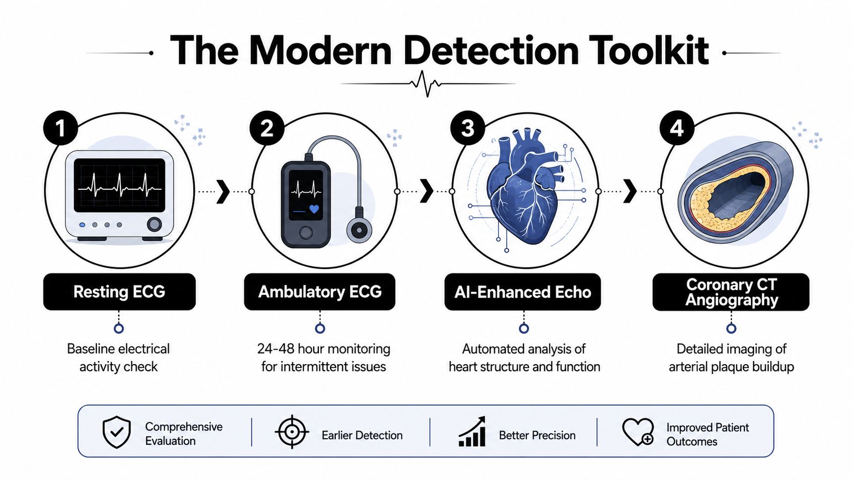
The easiest way to think about cardiac testing is to start with the question each test answers. A resting ECG looks at electrical patterns. An echocardiogram looks at structure and function. CT-based tools look for plaque. Cardiac MRI looks at tissue in greater detail. AI layers pattern recognition on top of those signals, which is why the modern toolkit feels less like one test and more like a diagnostic stack.
The Mayo Clinic notes that ECG-AI algorithms are being used in research to predict conditions including atrial fibrillation, amyloidosis, aortic stenosis, low ejection fraction, and hypertrophic cardiomyopathy Mayo Clinic. The American Heart Association has also noted that AI and machine learning applied to ECG can identify occult structural heart disease 1 to 2 years earlier than traditional testing Mayo Clinic summary of AHA perspective.
How the imaging pathway usually unfolds
The path often begins with a resting ECG, because it is quick, accessible, and useful for rhythm and conduction clues. If symptoms are intermittent, an ambulatory ECG adds time, which matters when a snapshot misses the event. Echocardiography follows when structure, valve function, or pumping performance needs a closer look.
From there, coronary calcium scoring can identify calcified plaque, and CT coronary angiography can show the arterial anatomy more directly. Cardiac MRI then adds tissue characterization, which is helpful when clinicians need to understand scar, inflammation, or muscle patterning. Readers comparing advanced MRI options can also look at what an AI full body MRI adds to integrated care, because the same principle applies, pattern recognition improves what human readers can already see.
A practical way to picture the hierarchy is this. ECG listens to electricity. Echo watches mechanics. CT maps the pipes. MRI studies the tissue. AI helps the reader detect subtleties that can be easy to overlook.
The attached process flow visual captures that progression well.
Detecting Disease in People Who Look Low-Risk
A fit runner can still carry hidden disease. That is the uncomfortable truth behind a lot of missed prevention. Routine risk calculators and standard labs are useful, but they can understate vulnerability in younger adults, especially when the person has inherited risk, vascular dysfunction, or a lifestyle profile that masks what is happening inside the vessel wall.
Where standard screening can miss the mark
The JACC review highlights the value of looking beyond traditional factors in young adults, including Lp(a), apoB, nonfasting lipids, and selected CAC scoring JACC review. That matters because many people in this group don't fit the old stereotype of the heart patient. They feel well, their office vitals look acceptable, and their first warning can arrive late.
Microvascular assessment is another frontier. Researchers at Helmholtz Munich and TUM developed fast raster-scan optoacoustic mesoscopy, or fast-RSOM, which can noninvasively capture three-dimensional capillary-level information through the skin and assess endothelial dysfunction at single-capillary and skin-layer resolution in humans Inside Precision Medicine. That matters because microvascular dysfunction can precede overt coronary disease.
A practical cardiology takeaway is simple. If the routine story says low risk but the biology feels off, deeper testing may be justified. That can include imaging, biomarker expansion, and rhythm analysis rather than relying on reassurance alone.
For patients who want broader, more accessible screening outside a specialty center, this discussion of whole-body MRI for integrated care fits the same logic, because the value comes from looking for hidden patterns before they become obvious disease.
A Tiered Screening Pathway for Precision Patients
A sensible prevention plan doesn't throw every test at every person. It escalates. That is especially important for medical tourists and concierge patients, because the goal is efficiency with clinical purpose, not testing for its own sake.
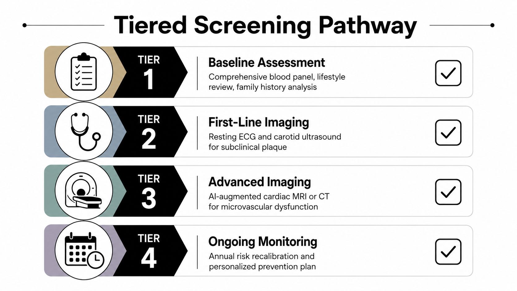
The four layers
Tier 1, Baseline assessment. Start with a thorough history, physical exam, blood pressure review, family history, and advanced biomarkers. The decision begins here, because good triage depends on good inputs.
Tier 2, First-line imaging. Resting ECG and echocardiography help identify rhythm, chamber size, valve problems, and obvious structural issues. If the question is “Is there already a visible abnormality?”, this is often where the first answer appears.
Tier 3, Advanced imaging. Coronary calcium scoring or CT coronary angiography comes next when the goal is plaque detection or anatomic clarification. A dedicated cardiac MRI can help when tissue characterization or functional detail is needed, and it can be paired with AI interpretation in specialized programs.
Tier 4, Ongoing monitoring. Annual recalibration keeps risk from becoming a static label. People change, biomarkers shift, and prevention plans should change with them.
The pathway used in a precision clinic may include modalities such as ECG, echocardiography, coronary CT, AI-augmented MRI, and cardiovascular biomarkers, which is why a coordinated center can compress what would otherwise take months into a single organized evaluation. One example in practice is the preventive heart screening pathway, where the value lies in sequencing, not in any one test.
How to know when to escalate
A patient with strong family history, high Lp(a), or discordant results deserves a lower threshold for advanced imaging. Someone with clean baseline data and no risk enhancers may stay in Tier 1 or Tier 2 longer. The decision is not about chasing certainty, it's about matching the intensity of testing to the intensity of risk.
Next Steps, Common Pitfalls, and Patient Questions
The biggest mistake is over-testing without a framework. The second is under-testing because a single normal result feels reassuring. A normal lipid panel, a clear ECG, or a good exercise tolerance test does not automatically close the book on coronary risk.
Common pitfalls to avoid
Treating one normal test as permanent clearance. Cardiac risk changes over time, so timing matters.
Using routine labs as a full verdict. They're useful, but they don't capture every high-risk pathway.
Escalating imaging without clinical context. More data is not always better if it doesn't answer a real question.
Ignoring data quality. When records are fragmented, interpretation gets sloppy, which is why an AI-powered healthcare data quality checker is relevant in modern care systems.
Questions patients often ask
How often should screening be repeated? It depends on baseline risk, age, symptoms, family history, and whether earlier tests showed abnormalities. Risk is dynamic, so the interval should be individualized.
Is AI-enhanced testing ready for routine use? It's already helping clinicians surface subtle findings earlier, especially in ECG interpretation and imaging review, but it works best as part of a supervised clinical pathway rather than as a standalone answer.
Do calcium scores matter in younger adults? They can, especially when the broader risk profile suggests disease may be starting earlier than expected. The result should be interpreted alongside biomarkers and history, not in isolation.
For readers who want a coordinated, physician-led evaluation, Longevity Medical Institute offers integrated cardiac assessment within a luxury medical setting, including advanced diagnostics that help clarify risk before symptoms begin. If you're ready to move from uncertainty to a structured prevention plan, visit Longevity Medical Institute to explore a personalized cardiac evaluation.
Short Disclaimer
This information is for educational purposes only and is not medical advice. It does not replace an evaluation by a qualified healthcare professional. For personalized guidance, please schedule a consultation.
Published July 29, 2026
Author
Dr. Kirk Sanford, DC, Founder & CEO, Longevity Medical Institute. Dr. Sanford focuses on patient education in regenerative and longevity medicine, translating complex therapies into clear, practical guidance for patients.
Medical Review
Dr. Félix Porras, MD, Medical Director, Longevity Medical Institute. Dr. Porras provides clinical oversight and medical review to help ensure accuracy, safety context, and alignment with current standards of care.
Last Reviewed: July 29, 2026