

Stem Cell Therapy for Brain Atrophy: Evidence and Options
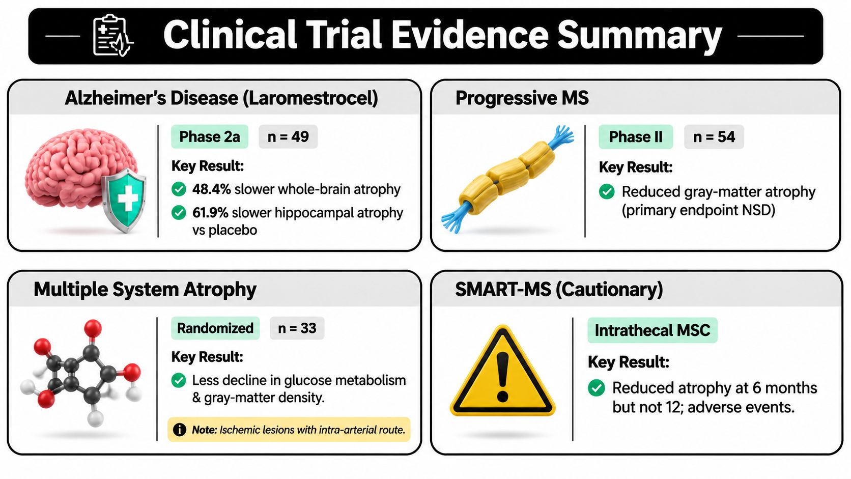
A 2025 phase 2a randomized, double-blind, placebo-controlled trial changed the tone of the conversation around Stem Cell Therapy for Brain Atrophy. In mild Alzheimer's disease, 49 participants received placebo or intravenous allogeneic bone-marrow-derived MSC therapy, and at 39 weeks the combined MSC groups showed 48.4% slower whole-brain volume decline and 61.9% slower left hippocampal volume decline versus placebo, with imaging changes correlated to cognitive testing rather than proving durable clinical benefit (PubMed trial report). For a field that has long been dominated by hope, that matters because it moves the discussion from vague regeneration claims to measurable MRI endpoints that can be tracked over time.
The practical takeaway is simple. Slower atrophy is not the same as reversing atrophy, but preservation of whole-brain and hippocampal volume could still be clinically important if it translates into better function over time. The study is still small, and it needs confirmation in larger trials, but it's one of the clearest signals yet that allogeneic MSCs may influence the biology of neurodegeneration in humans rather than just in theory (trial summary). If you want the broader Alzheimer's context, our related Alzheimer's article goes deeper into the same treatment family.
The 2025 Alzheimer's Trial That Changed the Conversation
The reason this trial stands out is that it used the right kind of endpoint for the right kind of question. Instead of asking only whether patients felt better, the investigators looked at whether IV allogeneic bone-marrow-derived mesenchymal stem cells, laromestrocel, could slow the structural loss that defines neurodegeneration. In the combined treated groups, the MRI signal was not subtle, 48.4% slower whole-brain decline and 61.9% slower hippocampal decline compared with placebo, with the effect measured against a disease where shrinkage is expected to continue (PubMed trial report).
Why that matters clinically
The hippocampus is one of the earliest structures tied to memory encoding and new learning, so when it shrinks, families often notice problems with short-term memory, orientation, and day-to-day recall. Whole-brain volume is broader, but it still tells you something important about the pace of neurodegeneration. In other words, these MRI findings matter because they point to tissue preservation, not just symptom management.
Practical rule: a positive biomarker signal is encouraging only if it survives larger trials and lines up with meaningful function, not just prettier scans.
What this trial did and didn't prove
The study used the modern language of brain-atrophy research, MRI-derived structural endpoints, not speculative claims about brain regrowth. It also fit a broader translational pattern in the field, where stem cell therapy is moving beyond isolated case reports and into repeated human testing across major neurodegenerative disorders, including Alzheimer's disease, Parkinson's disease, ALS, and Huntington's disease (2025 review of stem cell clinical trials). That broader review identified 94 trials in neurodegenerative diseases, which is an important marker of how much the field has expanded.
Still, the Alzheimer's result remains phase 2a evidence, not a finished answer. Small studies can overestimate benefit, and MRI improvement can outpace real-world function. The signal is real enough to pay attention to, but not strong enough to market as a proven treatment for brain atrophy.
Understanding Brain Atrophy and Why Volume Matters
A shrinking brain is not a single measurement, it is a pattern. Brain atrophy means loss of brain tissue over time. On MRI, that can appear as smaller hippocampi, thinner cortical regions, or enlarged ventricles, the fluid spaces that widen as surrounding tissue contracts. The pattern matters because different diseases affect different regions in different ways.
That is why MRI-based volume tracking is useful in brain-atrophy research. It gives clinicians a way to judge whether tissue loss is slowing, even before symptoms change in a way patients can feel. If you want a clearer explanation of how these scans are read in practice, this MRI results guide is a helpful reference.
Why the hippocampus and gray matter get so much attention
The hippocampus plays a central role in memory formation, while gray matter contains the neuronal cell bodies that support thinking, movement, and other higher functions. When these structures lose volume, cognition and function often decline as well, although the timing and pattern vary by disease. That is why researchers watch these regions closely when they test therapies for neurodegeneration.
Volumetric MRI shows change against a baseline scan. It is the same basic logic used in other serial measurements, where the value lies in whether a structure is holding steady or continuing to fall. The scan does not explain everything, but it gives a concrete marker for tissue preservation versus ongoing loss.
Diseases where atrophy matters most
Alzheimer's disease, progressive multiple sclerosis, and multiple system atrophy all involve accelerated tissue loss, even though the biology behind each condition is different. In Alzheimer's, memory-related structures are often affected early. In progressive MS, gray matter loss becomes increasingly tied to disability. In multiple system atrophy, structural and metabolic decline can move quickly, which is why investigators pay close attention to any therapy that may stabilize MRI markers.
Families sometimes first notice signs of cognitive overload before they recognize a neurological pattern. That does not diagnose brain atrophy, but it can help distinguish ordinary stress from more persistent changes in attention, memory, and mental stamina.
What the Clinical Trials Show
The Alzheimer's study deserves a close read because it tracked more than a single headline outcome. Investigators followed whole-brain volume, hippocampal volume, temporal cortex changes, ventricular enlargement, and MRI measures tied to neuroinflammation. That matters because atrophy rarely appears alone. It usually moves together with inflammation, microstructural disruption, and stress across connected networks.
The published report also linked changes in hippocampal and whole-brain atrophy with cognitive testing, which is encouraging. Still, a biomarker moving in the right direction does not prove treatment efficacy. A scan can improve while day-to-day function changes little, or changes only partly. That is why the field still needs larger, longer studies before anyone can claim the therapy alters disease course. See the original Alzheimer's trial report in PubMed.
Progressive multiple sclerosis
A randomized phase II study in 54 patients with progressive MS tested intrathecal MSC-derived neural progenitor cells. The primary clinical endpoint was not significantly different from placebo, yet MRI analysis found a reduced rate of gray-matter atrophy, especially in patients with more preserved gray matter at baseline. That pattern is interesting, but it is not a finished rule for selecting patients.
The practical reading is narrower. Earlier treatment may deserve more study because tissue that is still present may be easier to protect than tissue already lost. That is a biologically plausible hypothesis, not a proven clinical directive.
Multiple system atrophy
A randomized study in 33 patients with multiple system atrophy found that cerebral glucose metabolism and gray-matter density declined more in placebo-treated patients than in MSC-treated patients over 360 days. Those findings matter because they suggest the therapy may influence both structural and metabolic stability, not just one marker. The same study also showed that the intra-arterial component produced small ischemic lesions, a reminder that delivery route can change the risk profile in ways patients need to understand.
For readers who want a broader stem cell background, our mesenchymal stem cell research page places these clinical signals in a wider translational context.
How Mesenchymal Stem Cells May Support Brain Health
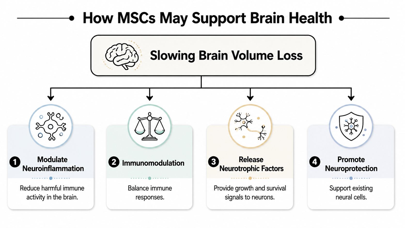
The main hypothesis is paracrine signaling, not direct cell replacement. MSCs are studied because they seem to release biologically active signals that can shape the brain's local environment, including BDNF, GDNF, and NGF described in preclinical and translational literature (histology-focused review). That is a much narrower claim than saying the cells rebuild lost cortex.
The main mechanisms under investigation
Researchers focus on neuroinflammation modulation, immunomodulation, neurotrophic signaling, growth factors, extracellular vesicles/exosomes, mitochondrial support, angiogenesis, synaptic protection, and reduction of oxidative stress. In practical terms, these mechanisms aim to make neurons and glia less vulnerable to ongoing injury and to preserve the microenvironment that supports tissue maintenance.
A recent histology-focused review found that stem cell therapies were associated with better neuron survival, improved synaptic structure, reduced gliosis, and partial restoration of tissue structure across neurodegenerative models, with effects varying by stem cell type, disease model, and delivery route (histology-focused review). That variability matters. Route selection and biologic potency are not minor details, they shape the technical result.
For patients who want the clinical framing, our mesenchymal stem cell therapy page explains how these mechanisms are considered in practice.
What this does not mean
Mechanistic plausibility is not proof of regeneration. MSCs are not established to regrow lost human brain tissue, and no honest clinician should present them that way. The evidence fits a neuroprotective and anti-inflammatory strategy, where the aim is to slow further deterioration rather than restore what has already disappeared.
Complementary Therapies for Brain Health
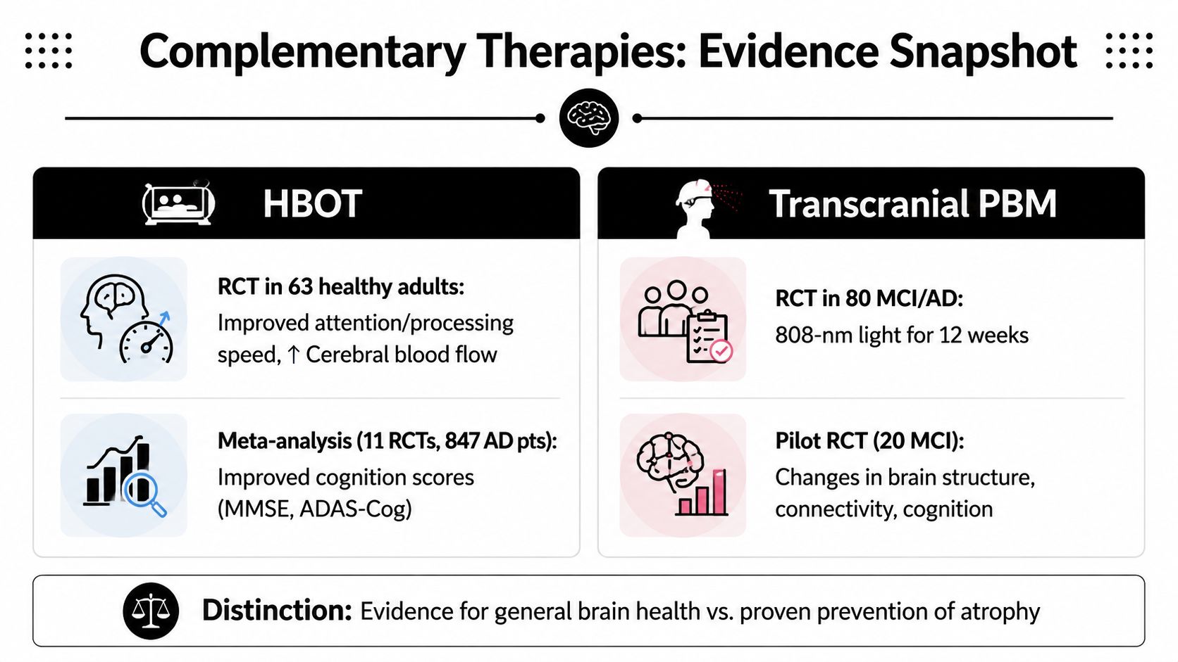
HBOT and transcranial PBM sit in a different evidence lane from MSCs, but they're relevant because both have human data tied to cognition and brain physiology. The key distinction is that brain-health support is not the same thing as proven prevention of atrophy. Patients often lump those together, and they shouldn't.
The HBOT literature is particularly useful when framed carefully. A randomized trial in 63 healthy adults over 64 found that a 3-month HBOT protocol significantly improved cognition, especially attention and processing speed, and MRI showed increased cerebral blood flow in several brain regions. A 2024 meta-analysis of 11 randomized trials involving 847 Alzheimer's patients also reported improvements in MMSE, ADAS-Cog, and activities of daily living (meta-analysis summary). The problem is not that the signals are absent, it's that the study quality and heterogeneity make broad conclusions hard.
Transcranial red and near-infrared PBM is arguably the more intriguing mechanistic partner. A 2026 randomized double-blind placebo-controlled trial of 80 people with MCI due to Alzheimer's disease used 808-nm PBM for 12 weeks, and MoCA improved 3.87 points versus a 0.74-point decline with placebo, with no device-related adverse events. A recent randomized pilot in MCI also reported changes in brain structure and functional connectivity after transcranial PBM, but that study had only 20 subjects, so it needs replication before anyone leans too hard on it.
Why the combination makes biological sense
The logic is coherent even if the evidence remains uneven. MSCs may support immunomodulation and trophic signaling, HBOT may improve oxygenation and perfusion, and PBM may influence mitochondrial/ATP signaling, neuroplasticity, and inflammation. That triad is conceptually attractive because it attacks different parts of the same problem, ongoing tissue stress.
At Longevity Medical Institute, hyperbaric oxygen therapy is one of several physician-supervised options sometimes considered alongside regenerative protocols, but it should still be viewed as supportive care rather than proof of atrophy reversal. For a closer look at that pairing, our HBOT and stem cell resource outlines the broader clinical context.
Neuroprotection Versus Neuroregeneration
How do clinicians separate preserving tissue from rebuilding it? In brain atrophy, that question shapes how I read the stem cell literature. Neuroprotection aims to slow additional damage, calm inflammatory stress, and preserve remaining tissue. Neuroregeneration would mean replacing tissue that has already been lost, and the current human evidence for MSCs in brain atrophy does not show that.
That is why I am cautious with “brain rejuvenation” claims. The better-supported interpretation is that stem cell therapy may help the brain resist ongoing injury, especially in selected early neurodegenerative disease, while MRI signals remain an imperfect proxy for real-world benefit. A smaller atrophy rate on imaging may be encouraging, but it does not yet prove lasting gains in cognition, independence, or quality of life.
The most defensible view is also the most practical one. Stem-cell therapy for brain atrophy appears more plausible as an anti-inflammatory and vascular-modulating strategy than as direct regeneration. That framing keeps expectations aligned with the evidence and helps patients judge whether a protocol is being offered as tissue preservation or as a promise to reverse established brain loss.
Patients should ask one question before anything else, “Is this being offered as tissue preservation, or as a promise to reverse established brain loss?” The difference changes how you interpret every claim.
Your Path to Personalized Brain Health Care
A serious brain-health evaluation starts with imaging, labs, and symptom history, not with a product pitch. At Longevity Medical Institute, that can include allogeneic stem cell therapies sourced from placental, Wharton's jelly, adipose, endometrial, and dental pulp programs, along with AI-enhanced full-body MRI, an in-house clinical laboratory measuring 120 biomarkers, and complementary therapies such as HBOT and photobiomodulation. Those tools help the team decide whether a patient is better suited to a neuroprotective plan, a recovery plan, or a different path entirely.
The practical sequence is straightforward. First comes a physician-led consultation, then a review of prior imaging and relevant medical history, then targeted testing to look for inflammatory, metabolic, vascular, and neurologic patterns that might influence treatment choice. If therapy is appropriate, the team then considers delivery route, follow-up timing, and what outcomes should be monitored over time.
For patients concerned about memory change, slowed processing, or MRI evidence of atrophy, the goal is not hype, it's precision. A good plan should tell you what the therapy might realistically do, what it probably won't do, and what needs to be watched closely after treatment.
If you're comparing options for brain health, recovery, or early neurodegenerative change, schedule a consultation with Longevity Medical Institute to review your imaging, symptoms, and candidacy for a personalized regenerative medicine plan. The right conversation starts with evidence, not promises, and a physician-guided assessment can help you decide whether stem cells, HBOT, PBM, or another approach makes the most sense for your situation.
Author
Dr. Kirk Sanford, DC, Founder & CEO, Longevity Medical Institute. Dr. Sanford focuses on patient education in regenerative and longevity medicine, translating complex therapies into clear, practical guidance for patients.
Medical Review
Dr. Félix Porras, MD, Medical Director, Longevity Medical Institute. Dr. Porras provides clinical oversight and medical review to help ensure accuracy, safety context, and alignment with current standards of care.
Last Reviewed: August 22, 2026
Short Disclaimer
This information is for educational purposes only and is not medical advice. It does not replace an evaluation by a qualified healthcare professional. For personalized guidance, please schedule a consultation.