

128-Slice Coronary CT: A Patient's Complete Guide
You're in a consultation room because something about your heart story hasn't felt fully settled. Maybe a stress test was borderline, maybe a parent had coronary disease early, or maybe a calcium score left you with more questions than answers. A 128-slice coronary CT is often the next step when the issue isn't just whether plaque exists, but whether the finding will change what happens next.
For many people, that's the key decision. A scan can be technically excellent and still be the wrong test if it won't clarify symptoms, guide treatment, or reduce uncertainty. The value of 128-slice coronary CT angiography is that it can help separate “watch this carefully” from “move to invasive angiography,” which is why it keeps showing up in modern cardiac workups.
That's also why the experience feels different from a generic “heart scan” conversation. People usually arrive at this test after a clinician wants a clearer view of the coronary arteries, especially when symptoms are stable, anatomy is still uncertain, or the next move depends on whether narrowing is present. A useful starting point is a preventive heart screening discussion, which you can review here.
When a Heart Scan Becomes the Next Question
A woman sits in clinic after a chest-pain workup that didn't give a simple answer. Her symptoms aren't dramatic, but they aren't ignorable either. Her clinician is weighing calcium scoring, a stress test, or a 128-slice coronary CT, and what matters most is not the label of the test, but whether it settles the question of coronary disease enough to guide the next step.
That's the right way to think about this scan. It's a decision tool, not a trophy image. If the question is, “Do these arteries have meaningful narrowing?” or “Do we need to escalate from monitoring to catheterization?”, coronary CT angiography can be very helpful, especially in stable patients whose symptoms or risk profile leave some ambiguity.
Who tends to land here
This test is commonly considered when chest symptoms are stable, family history is concerning, or earlier testing didn't line up cleanly with the clinical picture. It's also useful when clinicians want an anatomic look at plaque and stenosis without starting with an invasive catheter-based procedure. A broader preventive heart pathway can help frame that choice, and the concept of early detection is discussed in this overview.
The scan is not just about finding disease. It's about deciding whether disease is present in a way that matters enough to change treatment, intensify prevention, or move toward invasive angiography. That distinction is especially important when the patient is anxious, the symptoms are vague, or a calcium score suggests the arteries may be harder to interpret.
Practical rule: if the result won't change management, the scan may add information without adding clarity.
How 128-Slice Coronary CT Works
A heart that is beating, breathing, and shifting with every pulse is a difficult target. A 128-slice coronary CT handles that problem by collecting many thin cross-sections in rapid succession, then combining them into a three-dimensional dataset. That gives the cardiologist and radiologist several ways to inspect the coronary arteries, rather than relying on a single flat view.
Why the technical details matter
The key issue is not the slice count by itself. Technical references for 128-slice cardiac systems describe sub-millimeter isotropic volume acquisition, with slice acquisition commonly at 0.63 mm or smaller, at least 64 physical detector rows, and 128 slices per rotation for cardiac and vascular imaging, which supports sharper coronary lumen assessment and 3D reformations without directional blur (technical specification reference). In practical terms, that means the scanner can judge small coronary vessels, calcified plaque, and stents with less guesswork.
Rotation speed matters just as much. High-end 128-slice platforms pair that detector geometry with 0.35–0.39 s gantry rotation times, and some systems reach effective cardiac rotation times around 0.3 s, which helps freeze motion during the brief window when the heart is still enough to image well (technical specification reference). Temporal resolution works like camera shutter speed. The faster it is, the less blur comes from a moving heart.
What benchmark systems show
Published system specifications for Philips Ingenuity 128 list 64 detector rows, 128 slices per rotation, 0.4 s full-rotation speed, and 40 mm area coverage per rotation, while Siemens SOMATOM Perspective 128 lists 128 slices/rotation, 0.39 s rotation with iTRIM, and reconstructed slice widths down to 0.6 mm (Philips and Siemens specification summary). Those details affect how much of the heart can be covered with each heartbeat and how well the system tolerates motion.
A comparison study of 128-slice CT coronary angiography used 128 × 0.6 mm collimation, 0.3-second gantry rotation, and achieved 150 ms temporal resolution (PMC study). That kind of motion-freezing performance is what turns a spec sheet into images a clinician can use with confidence.
The reason this matters for care is simple. A scan is most useful when it changes the next step, whether that means reassurance, stronger preventive treatment, or referral for more invasive testing. In that sense, the technology is part of a broader pathway, and the decision point may be better understood through a practical overview of early heart disease detection.
The scanner's real job is simple. It has to capture a moving heart fast enough that the anatomy looks still.
What Happens During the Scan
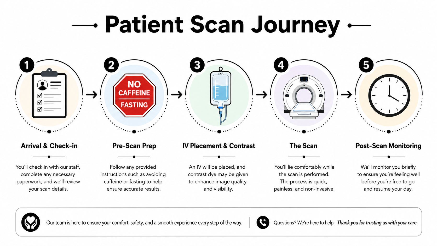
The day usually starts with a check-in, then a short pre-scan review. People are often asked to fast for several hours, avoid caffeine, and bring any relevant medication list, because heart rate and image timing can affect clarity. ECG electrodes are placed on the chest so the scanner can sync the image acquisition to the heartbeat.
The room, the machine, and the contrast
The scanner looks like a short tunnel with a table that slides through it. You'll lie still, hear the machine move, and usually hold your breath for a brief moment, often just a few seconds, while contrast is injected through an IV. Many patients feel a warm flush during contrast injection, which is common and usually passes quickly.
If the heart rate is fast or irregular, the team may use heart-rate control strategies before the scan, because steadier timing can improve image quality. One PubMed-indexed comparison showed that early 128-slice coronary CT validation performed well against invasive angiography, with 87% sensitivity, 97% specificity, 83% positive predictive value, and 97% negative predictive value at the segment level, though 58 of 879 segments (7%) were not assessable because of irregular rhythm, calcification, or tachycardia (PubMed study).
After the table slides back out
The visit doesn't end the moment the scan does. Staff usually monitor you briefly, then encourage hydration so the contrast clears more easily. The report is typically prepared later by the radiology team, and the scan is only as useful as the follow-up conversation that interprets it in the context of your symptoms and risk.
For a patient-facing overview of the wider MRI experience, including how imaging visits are organized, there's a helpful reference here.
How It Compares to Other Cardiac Imaging
The right test depends on the question. If the question is “How much calcium is in the coronaries?”, calcium scoring answers that without contrast. If the question is “Is there narrowing, plaque, or anatomy that explains symptoms?”, 128-slice coronary CT usually goes further because it visualizes the vessel lumen and the plaque around it.
| Test | Best For | Invasiveness | Typical Radiation | First-Line Role |
|---|---|---|---|---|
| 128-Slice CCTA | Coronary anatomy, stenosis, plaque, decision-making | Noninvasive | Low to moderate, protocol dependent | Often first-line in stable low-to-intermediate risk patients |
| Calcium scoring | Plaque burden and risk stratification | Noninvasive | Lower than contrast CT in many protocols | Screening and risk refinement |
| Invasive coronary angiography | Definitive lumen assessment and possible intervention | Invasive catheter-based test | Variable, procedure dependent | When anatomy or symptoms require direct catheter evaluation |
| Cardiac MRI | Function, tissue characterization, radiation-free imaging | Noninvasive | No ionizing radiation | Best when myocardial tissue questions matter more than coronary lumen detail |
| Lower-slice CT | Broader CT work, less detailed coronary imaging | Noninvasive | Protocol dependent | Less ideal for coronary detail |
A useful clinical comparison is that CCTA often acts as a gatekeeper. In stable chest pain, it can keep some patients away from unnecessary catheterization, while still sending the right patients onward when a lesion looks important.
Where the hierarchy changes
High coronary calcium complicates the picture because calcified plaque can blur the lumen and reduce specificity. In patients with advanced disease, the more useful question may be whether the anatomy is enough to explain the symptoms or whether functional testing is needed next. For a broader comparison with AI-enhanced imaging, see the clinic's overview of what is an AI full body MRI.
Clinical takeaway: the best test is the one that changes the next decision with the least risk.
Benefits, Limitations, and Real Risks
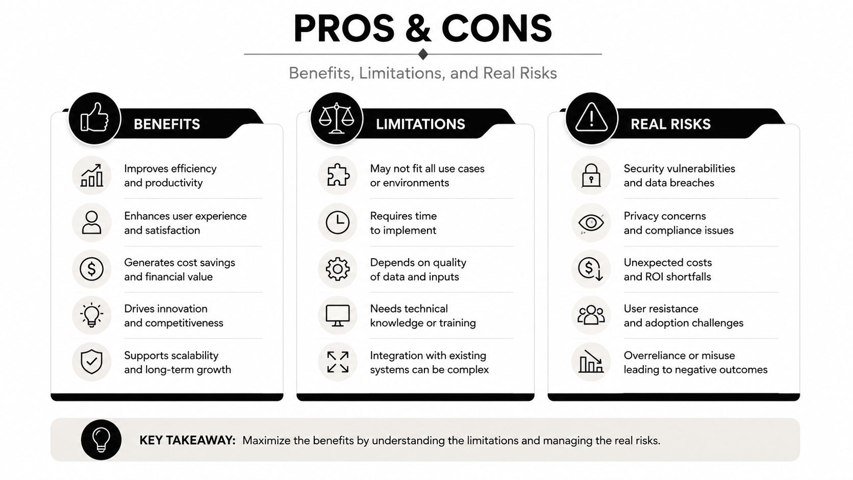
The strongest reason to choose 128-slice coronary CT is not that it produces a dramatic image. It is that, when the scan is technically good, it can help decide whether important coronary disease is present and whether the next step should be reassurance, prevention, more testing, or an invasive look at the arteries. A scan that shows plaque also does something practical, it gives the care team a clearer map of the problem. In a patient with vague chest symptoms, that map can separate “probably coronary” from “probably something else” and keep the workup focused.
What it does well
The best studies of coronary CT compared it with invasive angiography, which remains the anatomic reference when a catheter is already in the artery. Later protocol work showed that modern acquisition methods can lower radiation while preserving diagnostic usefulness. One randomized comparison of 128-slice dual-source CTCA found that the high-pitch spiral approach delivered 1.16 ± 0.60 mSv versus 3.82 ± 1.65 mSv for sequential CTCA, while specificity, PPV, and NPV stayed comparable in the analyzed group (PubMed radiation study). A separate dose review in the cardiac CT scan guide discusses how protocol choices, patient size, and heart rate all influence radiation in real practice. Another dose-focused report, in the context of coronary CT methodology, described lower exposure with prospectively triggered 128-MSCT and still acceptable image quality.
That matters because the value of the scan is not just the picture, it is whether the picture is good enough to change what happens next. A scan that is clear enough to rule out major obstruction can keep a patient away from an unnecessary catheter procedure. A scan that shows a suspicious lesion can justify moving on to functional testing or invasive angiography. A blurry scan that cannot answer the question is far less useful, even if the machine itself is advanced.
Where the trade-offs show up
Calcified plaque can create blooming artifacts, which makes calcium look thicker and can make a narrowing appear worse than it really is. Irregular rhythm can also reduce image quality because the scanner must time each picture to the heartbeat, much like a camera trying to freeze a moving hand. Contrast injection is another real trade-off, because iodinated contrast can stress the kidneys in susceptible patients or trigger allergic-type reactions. Whole-chest coverage can reveal incidental findings too, and that can start a chain of follow-up tests that may have little to do with the heart problem that brought the patient in.
That is why the scan should be judged as a decision tool, not as a trophy image. If the heart question is still unclear after the report, the next step may be a broader evaluation such as a comprehensive heart assessment, rather than assuming the CT alone settled the case. The scan helps most when it answers a specific question and changes the next clinical move.
A practical overview for patients is the cardiac CT scan guide. That kind of explanation is useful because the main downside is often not the scan itself, but the follow-up decisions the scan creates.
Reading the Results and What They Mean for Care
A report from 128-slice coronary CT usually comes down to whether there is narrowing, where it is, and how serious it appears. In published coronary CT practice, significant coronary stenosis is commonly defined as ≥50% luminal narrowing (PMC diagnostic threshold reference). That number matters because it gives the clinical team a starting point, not a complete answer.
Anatomy is not the same as function
A vessel can look narrowed on CT and still carry enough blood for the heart muscle. That is why an anatomic finding is only part of the decision, and why a moderate lesion often needs functional testing or cardiology review before anyone jumps to a procedure. A clean study, on the other hand, can be powerful reassurance because it makes major obstructive coronary disease much less likely.
The hardest calls are usually in people with high calcium or known coronary disease. In those cases, a CT may not just answer “is plaque there?”, it may help decide whether invasive angiography or revascularization is really warranted. That is the contrarian value of the scan, especially when the anatomy is noisy and the symptoms are not straightforward.
What to ask when the report is ambiguous
If a report shows borderline disease, ask whether the lesion matches the symptom pattern, whether calcium made the estimate less certain, and whether another test would change management. A broader heart assessment can help tie those threads together, and that framework is described here.
The right result is not the most dramatic one. It's the one that leads to the right next step.
Practical Considerations for Medical Tourists and Premium Clinics
Patients traveling for cardiac imaging should ask one question first. Is this a true 128-slice coronary CT platform with coronary protocol capability, or just a general CT labeled in a way that sounds similar? The distinction matters because cardiac performance depends on slice geometry, rotation speed, and ECG-gated workflow, not branding alone.
What a serious program should offer
Look for radiologist experience with coronary CTA, on-site cardiology review, and a report pathway that doesn't leave you waiting in another time zone for answers. It also helps if the clinic can coordinate calcium scoring, advanced lipid testing, and broader cardiovascular assessment in one visit, because coronary CT is most useful when it sits inside a bigger risk picture.
What to verify before you book
The clinic should be able to explain whether the scanner supports sub-millimeter coronary acquisition, how the report is reviewed, and when you'll get a result. Ask whether follow-up can be done remotely, whether the archive is portable, and whether the imaging is integrated with a broader prevention plan rather than treated as an isolated scan.
Longevity Medical Institute offers a 128-slice coronary CT as part of a preventive heart evaluation, alongside coronary calcium scoring and broader cardiovascular assessment. For some travelers, that kind of integrated workflow is the reason the trip is worth making. A premium clinic setting is only useful if the imaging is clinically solid and the follow-up is organized.
Decision Framework and Common Patient Questions
If symptoms are stable and the question is coronary disease, 128-slice CCTA is often the first-line test. If calcium is very high or known coronary disease is already established, pairing CT with functional imaging often makes more sense. If the CT result is ambiguous and management still hangs in the balance, invasive angiography becomes the next conversation.
Common questions patients ask
How much radiation is it, really? It depends on the protocol. As noted earlier, modern 128-slice approaches can bring dose down meaningfully, but the exact number varies with technique and patient factors.
Is it safe if my kidneys are not perfect? That depends on your kidney function, hydration status, and whether contrast is necessary. The contrast decision should be individualized, not automatic.
Can it be repeated? Sometimes, yes, but only if the result will change care. Repeating a scan because the first one was non-diagnostic is different from repeating it out of routine.
Is it for people without symptoms? Sometimes in selected high-risk people, but not as a casual screening test. A strong family history alone doesn't automatically mean the scan is the right first step.
Bottom line: the scan is most valuable when it answers a decision, not when it merely produces an image.
Author
Dr. Kirk Sanford, DC, Founder & CEO, Longevity Medical Institute. Dr. Sanford focuses on patient education in regenerative and longevity medicine, translating complex therapies into clear, practical guidance for patients.
Medical Review
Dr. Félix Porras, MD, Medical Director, Longevity Medical Institute. Dr. Porras provides clinical oversight and medical review to help ensure accuracy, safety context, and alignment with current standards of care.
Last Reviewed: August 1, 2026
Short Disclaimer
This information is for educational purposes only and is not medical advice. It does not replace an evaluation by a qualified healthcare professional. For personalized guidance, please schedule a consultation.
If you're trying to decide whether a 128-slice coronary CT makes sense for your symptoms, calcium score, or family history, Longevity Medical Institute can help you interpret the next step with physician-led cardiovascular assessment and advanced imaging. Visit Longevity Medical Institute to review preventive heart screening options and arrange a consultation.